Figures & data
Figure 1 Antiproliferative effects of niclosamide in human colon cancer cells.
Notes: Colon cancer cell lines (A) SW620, (B) HCT116, and (C) HT29 were treated with different concentrations (60, 12, 2.4, 0.48, and 0.098 µM) of nifuroxazide (nifu), niclosamide (nicl), cryptotanshinone (cry), and a lantolactone (ala), respectively, for 72 h. Cell proliferation in each group was detected by MTT assay. (D) HCT116, SW620, and HT29 cell lines were suppressed with niclosamide treatment at different concentrations (20, 10, 5, 2.5, and 1.25 µM) for 72 h. Subsequently, cell proliferation in each group was detected by MTT assay. The data were obtained from 3 independent experiments.
Abbreviation: IC50, half maximal inhibitory concentration.
Abbreviation: IC50, half maximal inhibitory concentration.
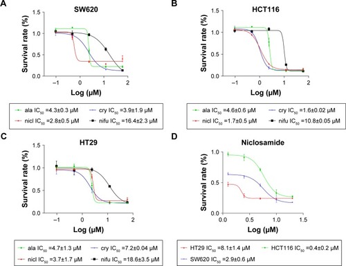
Figure 2 MD analysis of the effect of niclosamide on the activity cavity of STAT3.
Notes: (A) Molecular structure of niclosamide. (B) Last snapshot of STAT3/niclosamide in 50 ns MD simulations.
Abbreviation: MD, molecular dynamics.
Abbreviation: MD, molecular dynamics.
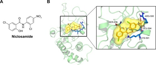
Figure 3 Inhibition of STAT3 phosphorylation by niclosamide in colon cancer cells HCT116 and SW620.
Notes: (A) SW620 cells were treated with niclosamide (5 µM) for different lengths of time (0, 2, 4, 6, 8, and 12 h). Total protein was extracted, and the expression levels of P-STAT3, STAT3, and GAPDH proteins were detected by Western blot analysis. (B) HCT116 cells were treated with niclosamide (5 µM) for different lengths of time (0, 2, 4, 6, 8, and 12 h). Total protein was extracted, and the expression levels of P-STAT3, STAT3, and GAPDH proteins were detected by Western blot analysis. (C) SW620 cells were treated with niclosamide at different concentrations (0.5, 1, 2.5, 5, and 10 µM) or vehicle control (DMSO) for 12 h. (D) HCT116 cells were treated with niclosamide at different concentrations (0.5, 1, 2.5, 5, and 10 µM) or vehicle control (DMSO) for 12 h. Total protein was then extracted and detected by Western blot analysis. The data were obtained from 3 independent experiments. *P<0.05.
Abbreviation: DMSO, dimethyl sulfoxide.
Abbreviation: DMSO, dimethyl sulfoxide.
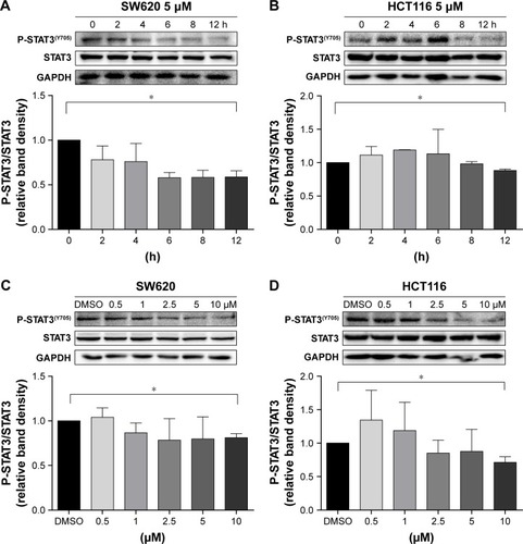
Figure 4 Sensitization of colon cancer cells to erlotinib by niclosamide.
Notes: (A) HCT116, SW620, and HT29 cells were seeded in a 96-well plate at a density of 5,000 cells per well and then cultured for 72 h. The cells were treated with erlotinib and gefitinib at indicated concentrations (60, 12, 2.4, 0.48, and 0.096 µM). After the 72 h treatment, cell proliferation in each group was detected by MTT assay. (B) HCT116, SW620, and HT29 cells were seeded at a density of 5,000 cells per well. Cells were treated with erlotinib at various concentrations (60, 12, 2.4, 0.48, and 0.096 µM) or/and niclosamide (0.5 and 1 µM) in triplicate for 72 h to detect the effects of combined erlotinib and niclosamide by MTT assay. (C) HCT116, SW620, and HT29 cells were treated with niclosamide (0.5 and 1 µM) in triplicate for 72 h to detect the effects of niclosamide by MTT assay. (D and E) For colony formation assay, SW620 cells were used. The number of colonies was counted after SW620 cells were incubated and treated with erlotinib (10 µM) or/and niclosamide (2.5 µM) for 1 week and then stained with crystal violet. *P<0.05.
Abbreviations: DMSO, dimethyl sulfoxide; E, erlotinib, nicl, niclosamide.
Abbreviations: DMSO, dimethyl sulfoxide; E, erlotinib, nicl, niclosamide.
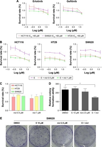
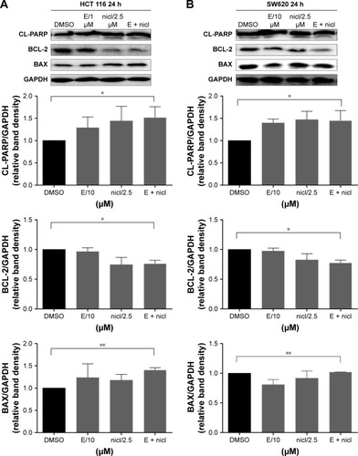
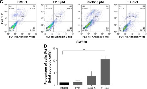
Figure 5 Induction of apoptosis in colon cancer cells by combined niclosamide and erlotinib.
Notes: (A) HCT116 cells were treated with erlotinib (10 µM) and/or with or without niclosamide (2.5 µM) for 24 h. P-STAT3, BCL-2, and BAX were detected by Western blot analysis. (B) SW620 cells were treated with erlotinib (10 µM) and/or with or without niclosamide (2.5 µM) for 24 h. P-STAT3, BCL-2, and BAX were detected by Western blot analysis. (C and D) SW620 cells were treated with erlotinib (10 µM) and/or with or without niclosamide (2.5 µM) for 24 h. The level of apoptosis was evaluated with Annexin V and PI, followed by detection using flow cytometry. *P<0.05; **P<0.01.
Abbreviations: DMSO, dimethyl sulfoxide; E, erlotinib, nicl, niclosamide.
Abbreviations: DMSO, dimethyl sulfoxide; E, erlotinib, nicl, niclosamide.