Figures & data
Figure 1 Migration of HepG2 cells was promoted by chemotherapy-induced apoptosis. After the cells on the bottom of the wells being treated with 2.5 μM VP-16, crystal violet staining assay was applied to analyze the influence of the treated cells on the migration of HepG2 cells from day 1 to day 7. Results were expressed as the mean ± SD of three independent experiments, *p < 0.05.
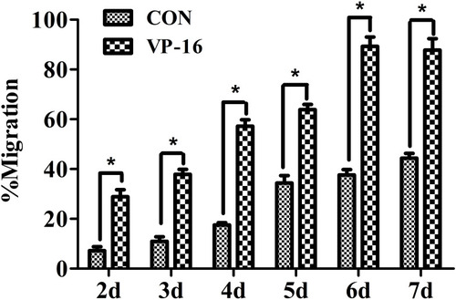
Figure 2 Berberine reversed the apoptosis-induced migration of HepG2 cells. (A) After the cells on the bottom of the wells being treated with 2.5 μM VP-16, 3.125 μM Berberine was applied to the system and cultured for 6 days. Crystal violet staining assay was applied to analyze the migration of HepG2 cells. Cell numbers were counted under the microscope. (B) Cells from the outer side of the membrane were photographed under the microscope. (C) The cells were then exposed to the culture medium of the 7th day’s incubation for the Scratch-wound assay. (D) Migration of the cells was analyzed by Image J. Each bar represents the mean ± SD of three independent experiments, n=3, *p < 0.05.
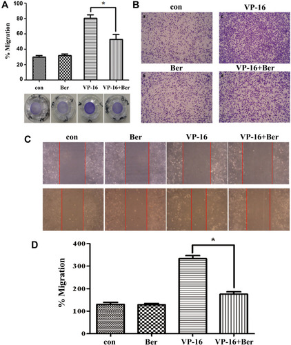
Figure 3 Berberine reversed the apoptosis-induced Adhesion of HepG2 cells. (A) Cells were cultured in the culture medium collected from the 7th day’s incubation and shaken for 2 h at 37°C. (B) Cells in culture medium collected from the 7th day’s incubation were added to the FN-coated wells, and allowed to attach for 2 h. Attached cells were fixed, stained with 4% SRB. (C) stained cells were lysed in 10mM Tris-base and the released stain was quantified by absorbance at a wavelength of 540 nm. Each bar represents the mean ± SD of three independent experiments, n=3, *p < 0.05.
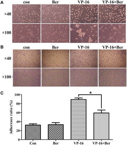
Figure 4 The protein expression of iPLA2 and LOX-5 in HepG2 cells treated by VP-16 and/or Berberine. (A) The cells after 5 days’ incubation were collected and the protein expression of iPLA2 and LOX-5 in HepG2 cells were measured by Western blot analysis. (B) Relative quantification of iPLA2 levels expressed relative to control. (C) Relative quantification of LOX-5 levels expressed relative to control. (D) The LTB4 levels in HepG2 cells treated by VP-16 and/or Berberine. The supernatants of each group were collected each day during the 7 days’ incubation. The LTB4 levels were measured by ELISA. Each bar represents the mean ± SD of three independent experiments, n=3, *p < 0.05 compared with control; **p < 0.01 compared with control; #p < 0.05 compared with VP-16 group. ##p < 0.01 compared with VP-16 group.
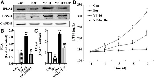
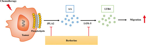
Figure 5 Schematic illustration of the mechanism by which Berberine inhibits the apoptosis-induced metastasis by suppressing the AA-LOX-5 pathway. Chemotherapeutics-induced tumor cell apoptosis can activate the LOX pathway, and subsequently release inflammatory factor LTB4 which ultimately stimulates the adhesion and migration of the small number of surviving tumor cells. And Berberine can reverse the adhesion and migration by inhibiting the expression of iPLA2 and LOX-5 and reduce LTB4 production in the tumor microenvironment.