Figures & data
Figure 1 RSF-1 was upregulated in cervical cancer tissues and decreased after effective treatment. (A) The expression of RSF-1 mRNA in cervical cancer tissue and normal control from the ONCOMINE database. (B) The expression of RSF-1 mRNA and (C) RSF-1 protein in 16 pairs of tumor samples from cervical cancer patients before and after radiochemotherapy were examined by qRT-PCR and Western blot.
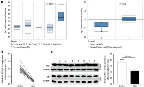
Figure 2 RSF-1 siRNA confers sensitivity to radiation in HeLa and SiHa cells. HeLa and SiHa cells transfected with siRNA-NC or siRNA-RSF-1 were irradiated with 0, 4 and 10Gy and then subjected to (A) CCK-8 assays and (B) colony formation assays. HeLa and SiHa cells transfected with pcDNA3.1-NC or pcDNA3.1-RSF-1, were irradiated with 0, 4 and 10Gy and then subjected to (C) CCK-8 assays and (D) colony formation assays. Data were presented as mean ± SD from three independent experiments. **P<0.01, *P <0.05 as compared with the siRNA-NC or pcDNA3.1-NC groups.
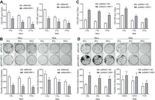
Figure 3 RSF-1 siRNA promotes radiation-induced cell cycle redistribution and apoptosis. HeLa and SiHa cells transfected with siRNA-NC or siRNA-RSF-1 were irradiated with 0, 4 and 10Gy and then subjected to (A) cell apoptosis and (B) cell cycle analysis. Data were presented as mean ± SD from three independent experiments. ***P<0.001, **P<0.01, *P <0.05 as compared with the siRNA-NC group.
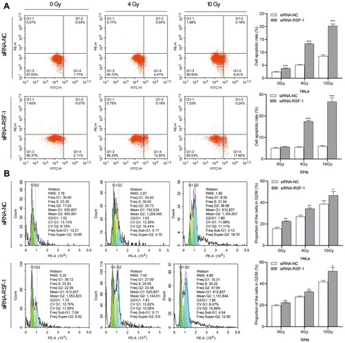
Figure 4 RSF-1 siRNA promotes radiation-induced DNA damage. (A) The extent of DNA damage was assessed by Comet assay. The percentage of DNA in comet tail (Tail DNA %) was quantified and graphed for each group. (B) The expression of H2AX and γH2AX proteins were examined by Western blot. The ratio of γH2AX level relative to H2AX after normalization to GAPDH was quantified and graphed for each group. (C) HeLa cells transfected with siRNA-NC or siRNA-RSF-1 were irradiated with 4Gy and then stained with an anti-RSF-1 (green) or an anti-γH2AX (red) antibody. RSF-1- and γH2AX-positively stained cells were scored and showed in right panel. The experiments were repeated three times independently. ***P<0.001, **P<0.01, *P <0.05 as compared with the siRNA-NC group.
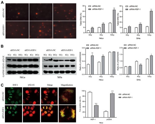
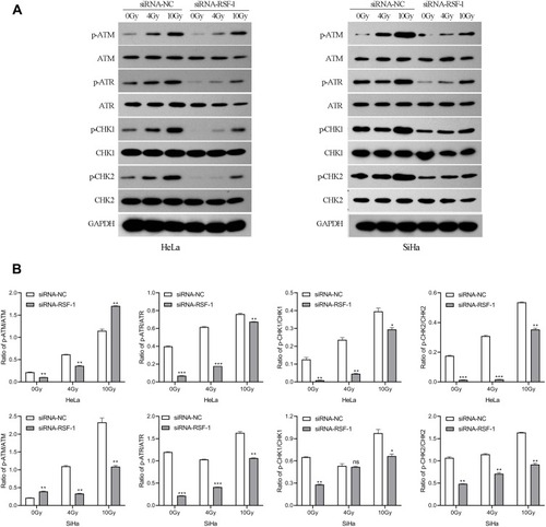
Figure 5 RSF-1 siRNA impairs ATM/ATR signaling pathway activated by radiation. (A) HeLa and SiHa cells transfected with siRNA-NC or siRNA-RSF-1 were irradiated with 0, 4 and 10Gy, and the ATM/ATR pathway proteins were examined by Western blot. (B) The relative gray values of p-ATM, p-ATR, p-CHK1 and p-CHK2 were calculated and graphed for each group. The experiments were repeated three times independently. ***P<0.01, **P<0.01, *P <0.05 as compared with the siRNA-NC group.