
Figures & data
Table 1 IC50 Value of Cisplatin and Calycosin in Twelve GC Cell Lines
Figure 1 Cytotoxic effects of calycosin on GC cells. (A) Twelve human GC cell lines (AGS, KATO-3, MKN-28, MKN-45, NCI-N87, SNU-5, SNU-216, SNU-484, SNU-668, YCC-1, YCC-6, and YCC-16) were treated with different concentrations of calycosin and cisplatin for 24 h, and the cell cytotoxic effects were determined by the CCK-8 assay. (B) Four human normal cell lines (GES-1, IMR-90, L-02, and 293T) were treated with different concentrations of calycosin and cisplatin for 24 h, and the cell cytotoxic effects were determined by the CCK-8 assay. (C) Twelve human GC cell lines were treated with the IC50 of calycosin and cisplatin for 0, 3, 6, 12, 24, and 48 h, and the cell cytotoxic effects were determined by the CCK-8 assay. (D) Four human normal cell lines were treated with the IC50 of calycosin and cisplatin for 0, 3, 6, 12, 24, and 48 h, and the cell cytotoxic effects were determined by the CCK-8 assay. Representative data from at least three independent tests. *p < 0.05, **p < 0.01, and ***p < 0.001.
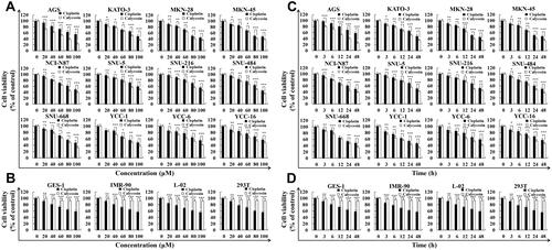
Figure 2 Calycosin induces apoptosis on AGS cells. (A) AGS cells were treated with 47 μM calycosin (0, 3, 6, 12, and 24 h) and stained with Hoechst 33342/PI. Fluorescence microscopy of apoptotic AGS cells (original magnification: 400×). (B) AGS cells were stained with Annexin V-FITC/PI and analyzed by flow cytometry. (C) AGS cells were labeled with the JC-1 fluorescent probe and analyzed by flow cytometry. (D) Western blot analysis of the expression levels of Bad, Bcl-2, cle-caspase-3, cle-PARP, and cytochrome C in AGS cells treated with calycosin for different times (0, 3, 6, 12, and 24 h). Representative images from at least three independent tests. *p < 0.05, **p < 0.01, and ***p < 0.001.
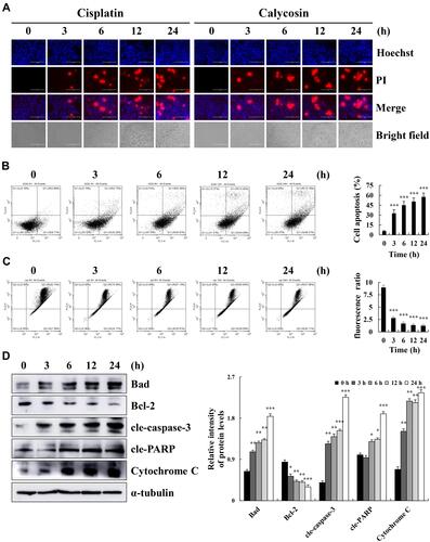
Figure 3 Calycosin affects the expression of MAPK, STAT3, and NF-κB pathway-related proteins in AGS cells. (A) AGS cells were treated with calycosin (0, 3, 6, 12, and 24 h). The expression levels of p-ERK, p-p38, p-JNK, p-STAT3, NF-κB, and IκB-α were detected by Western blotting; α-tubulin was used as the internal control. (B) The expression levels of nuclear STAT3, NF-κB, and IκB-α were detected by Western blotting, and Lamin B1 was used as a nuclear loading control. (C) AGS cells were pretreated with FR180204 (12.1 μmol/L) for 30 min, and the expression levels of p-ERK, ERK, p-STAT3, STAT3 and cle-caspase-3 were detected by Western blotting. (D) AGS cells were pretreated with SB203580 (12.1 μmol/L) for 30 min, and the expression levels of p-p38, p38, p-STAT3, STAT3 and cle-caspase-3 were detected by Western blotting. (E) AGS cells were pretreated with SP600125 (12.1 μmol/L) for 30 min, and the expression levels of p-JNK, JNK, p-STAT3, STAT3 and cle-caspase-3 were detected by Western blotting. Representative data from at least three independent tests. *p < 0.05, **p < 0.01, and ***p < 0.001.
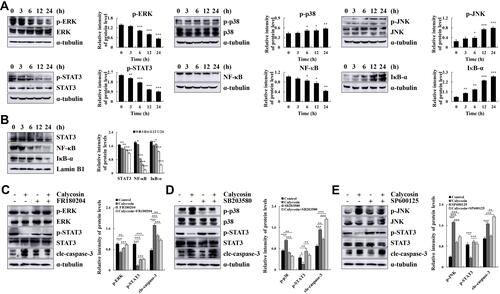
Figure 4 The effect of calycosin on intracellular ROS levels in AGS cells. (A) AGS cells were treated with calycosin (47 μM), and ROS generation was analyzed by flow cytometry. (B) The apoptosis of AGS cells was analyzed after pretreatment with NAC by flow cytometry. (C) Western blotting was used to analyze the expression of p-ERK, p-p38, p-JNK, p-STAT3, NF-κB, IκB-α, cle-caspase-3, cle-PARP, and cytochrome C protein; α-tubulin was used as the internal control. (D) Nuclear STAT3, NF-κB, and IκB-α expression levels were determined by Western blotting; Lamin B1 was used as a nuclear loading control. Representative data from at least three independent tests. *p < 0.05, **p < 0.01, and ***p < 0.001.
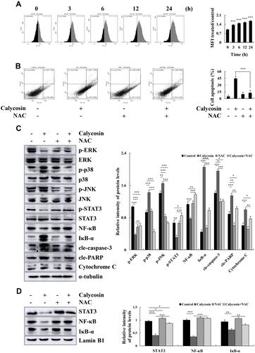
Figure 5 Effects of calycosin on the cell cycle and the expression of cell cycle-related proteins in AGS cells. (A) AGS cells were treated with 47 μM calycosin for 0, 3, 6, 12, and 24 h, and cell cycle distribution was analyzed by flow cytometry. (B) The expression levels of G0/G1 cell cycle proteins CDK2, CDK4, CDK6, cyclin D1, cyclin E, p21, and p27 were examined by Western blotting; α-tubulin was used as the internal control. (C) The cell cycle of AGS cells was analyzed after pretreatment with NAC by flow cytometry. (D) The expression level of cell cycle was analyzed by Western blotting after pretreatment with NAC; α-tubulin was used as the internal control. Representative data from at least three independent tests. *p < 0.05, **p < 0.01, and ***p < 0.001.
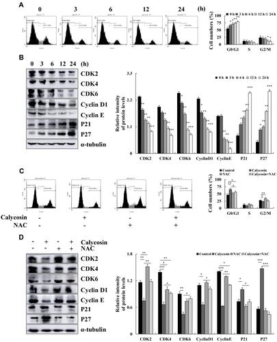
Figure 6 Effect of calycosin on the migration of AGS cells. (A) AGS cells were treated with calycosin (47 μM), and the images were taken under an inverted microscope. (B) Western blotting was used to analyze the expression of SNAI 1, E-cadherin, and β-catenin. (C) AGS cells were pretreated with NAC, followed by the addition of calycosin (47 μM) and imaging under an inverted microscope. (D) The expression of SNAI 1, E-cadherin, and β-catenin was analyzed after pretreatment with NAC; α-tubulin was used as the internal control. Representative data from at least three independent tests. *p < 0.05, **p < 0.01, and ***p < 0.001.
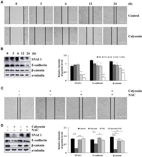
Figure 7 Schematic illustration of the underlying mechanism of calycosin effects on signaling pathways and the induction of apoptosis in GC cells.
