Figures & data
Figure 1 Chest computed tomography revealed the different stages of the patient’s lung tumor. (A) Initial diagnosis revealed a nodular high-density shadow in the posterior segment of the upper lobe tip of the left lung in April 2020. (B) CT scan after the surgery in June 2020.
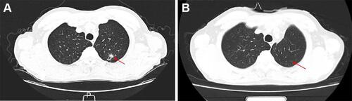
Figure 2 Next-generation sequencing findings of LOC101927967-ALK fusion. (A) The Integrative Genomics Viewer snapshot of LOC101927967-ALK. (B) Schematic representation of the LOC101927967 intergenic region ALK fusion, this variant was generated by the fusion of intergenic region between LOC101927967 and REG3G with exons 20–29 of ALK.
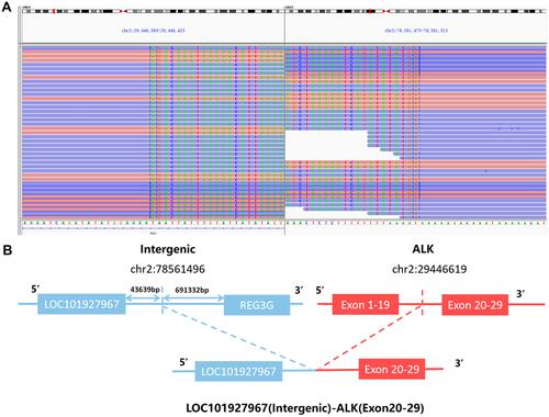
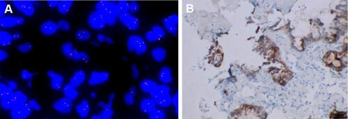
Figure 3 LOC101927967-ALK fusion in the patient with lung adenocarcinoma. (A) A split signal of ALK was observed with a frequency of 66% in the fluorescence in situ hybridization image. (B) The immunohistochemistry staining indicated strong positive ALK protein expression.