
Figures & data
Figure 1 Computed tomographic imaging: The enhanced computed tomographic (CT) scan revealed a mass of 30×40 mm in the upper pole of left renal that was classified as Bosniak category IV.
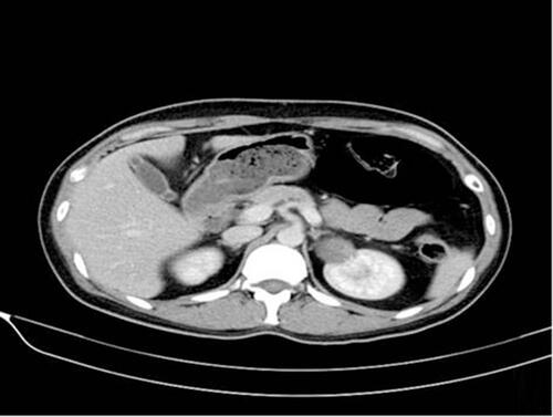
Figure 2 Pathological features of the renal carcinoid: (A) H&E (×100) showing nests and cords of neuroendocrine cells, which were arranged in a rosette like structure; (B) Positive immunostaining in tumor cells with synaptophysin (×200); (C) Intense positive immunostaining for chromogranin in tumor cells (×100); (D) Positive immunostaining in tumor cells with CD56 (×200); (E) Positive immunostaining in tumor cells with neuron specific enolase (×100).

Table 1 Clinical Features of the Patients
Table 2 Pathologic Features of the Patients
Table 3 Immunohistochemical Features of the Patients