Figures & data
Figure 1 A regular-shaped mass in the right breast in the ultrasound test.
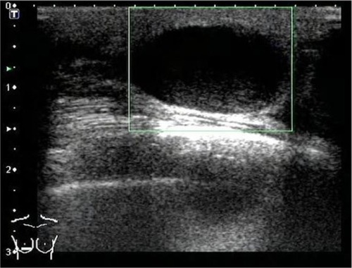
Figure 2 HE staining showing tumor cells with CD68 and vimentin-positive expression (×200 and ×400).
Abbreviation: HE, hematoxylin–eosin.
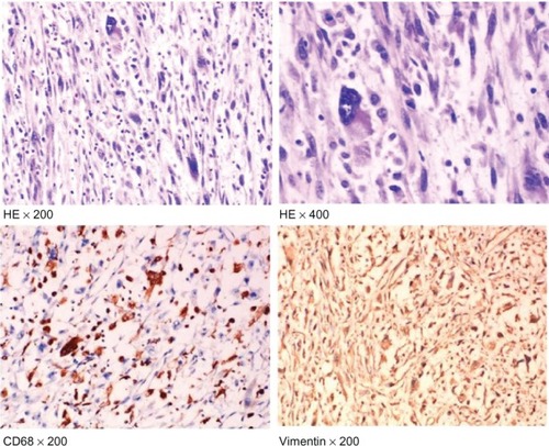
Figure 3 Enhanced computed tomography scanning showed lung metastases.
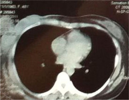
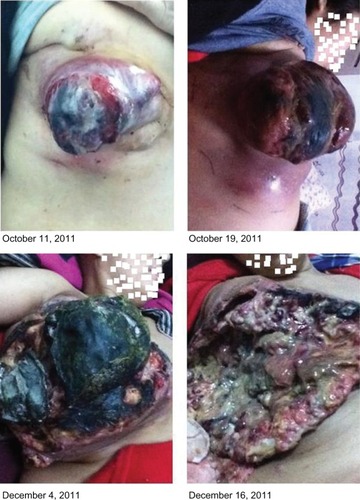
Figure 4 Tumor images after surgery.