Figures & data
Figure 1 Tumor cells containing large amounts of eosinophilic cytoplasm in the interstitium of vaginal squamous epithelium (squamous epithelium lacking lesions) presents a pattern of nodular, nested, single cell growth with central cystic degeneration. There is a large amount of eosinophilic basement membrane-like material surrounding the tumor cells.
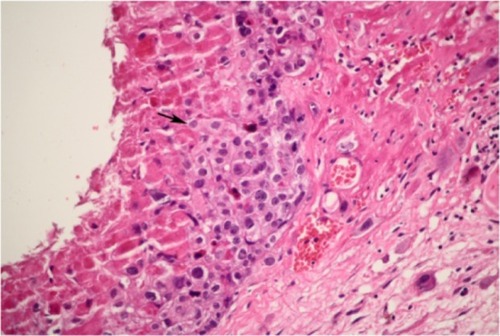
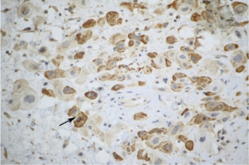
Figure 2 Immunohistochemical staining showed that the tumor cells were positive for human chorionic gonadotropin (hCG) (original magnification ×150).
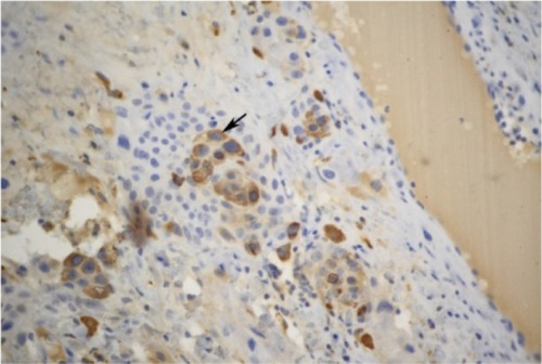
Figure 3 Immunohistochemical staining showed that the tumor cells were positive for human placental lactogen (hPL) (original magnification ×300). All the cells stained as claybank by 3,3’-diaminobenzidine chromagen (DAB).