Figures & data
Figure 1 Clinical characteristics of proband (V-4). (A) ECG showed sinus bradycardia and occasional sinus cardiac arrest. (B) 24 hr ambulatory electrocardiogram showed the changes in heart rate and sinus cardiac arrest in a day. (C) EMG displayed spontaneous electromyographic activity in limb muscles. (D) Muscular MRI showed mild fatty infiltration in the skeletal muscle. (E) The patient (proband) developed without any obvious symptoms of neuromuscular disease.
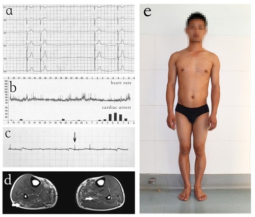
Figure 2 Family Pedigree. All individuals in the pedigree are identified by their Roman numerals below the symbol. Arabic numerals denote each individual in a generation. Squares represent males, and circles represent females. The proband (V-4) is designated with an arrow. Open symbols are unaffected individuals, filled symbols are hemizygous individuals, symbols with backslashes are heterozygous individuals, and symbols with diagonal lines indicate deceased subjects. Underlined numerals are obligate carriers who did not undergo molecular analysis. Numeral with a question mark (?) is an individual with an uncertain genetic status.
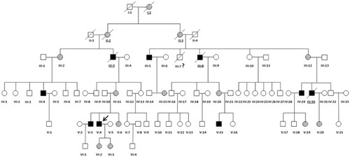
Table 1 Clinical Features Of The Proband And 9 Affected Family Members
Figure 3 Sequencing results of EMD mutation. (A) Image a shows results for a normal individual. Sanger sequencing demonstrated a duplication mutation (c.405dup) in exon 5 of EMD in the hemizygous individual (B) and the overlapping peaks in the heterozygous individual (C).
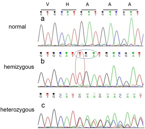
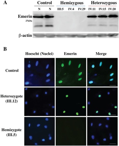
Figure 4 Immunoblot and immunohistochemistry results revealed that EMD hemizygous individuals do not express emerin protein. (A) Immunoblotting of emerin protein (29 kDa) expression from total protein extracts of lymphocytes from the hemizygotes (III5, IV4, IV29), heterozygotes (IV11, IV15, IV20) and a normal control, with use of the emerin monoclonal antibody. No emerin protein bands were detected in the hemizygous patients. (B) Left panels, nucleus (Hoechst) staining (blue); middle panels, emerin staining (green); right panels, merged emerin and nucleus staining. Patients include a normal male control, heterozygote female (III.12) and a male hemizygote (III.5). Scale bar represents 50 μm.