
Figures & data
Figure 1 Radiographic (MRI) images and histology of GBMs.
Notes: (A) shows an MRI of a GBM before (left) and after (right) surgery. Yellow circles show the area of tumor (left) and the resection cavity following surgery (right). (B) (low power, 100×) and (C) (high power, 600×) are hematoxylin/eosin stains of a section of a GBM used in histopathologic diagnosis. (B) shows the typical hypercellularity, cytological atypia, and prominent pseudopalisading necrosis of a GBM. (C) at higher power (same tumor, different section), better illustrates the cellular atypia and mitotic activity in the GBM.
Abbreviations: GBM, glioblastoma multiforme; MRI, magnetic resonance imaging.
Abbreviations: GBM, glioblastoma multiforme; MRI, magnetic resonance imaging.
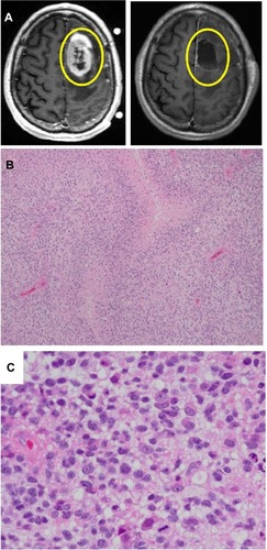
Figure 2 Two main modes of EV formation.
Notes: (A) shows the exosome pathway, whereby materials are taken from the cell surface into the endosomal system, with later invaginations forming MVB. If the MVB fuses with the plasma membrane, it releases the internal vesicles into the extracellular space as exosomes. (B) shows the formation of microvesicles as shed vesicles, budding directly off from the plasma membrane.
Abbreviations: EV, extracellular vesicle; MVB, multivesicular body.
Abbreviations: EV, extracellular vesicle; MVB, multivesicular body.
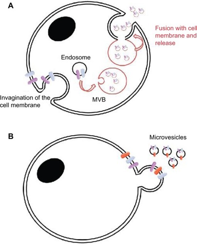
Table 1 Subclassifications of glioblastomas (based on TCGA)Citation25