Figures & data
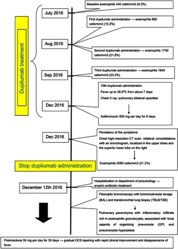
Figure 1 Chest x-ray: bilateral pulmonary opacities.
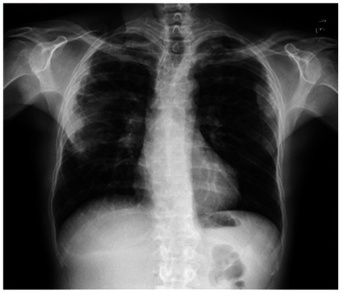
Figure 2 Chest high-resolution CT scan pre-post oral corticosteroids (OCS) treatment: (A) bilaterally, areas of parenchymal thickening with plane bronchograms, confluent, located in the upper lobes bilaterally, and peripherally, in the postero-lateral mantle area of the supero-lateral segment of the upper lobe. (B) Areas of parenchymal thickening with plane bronchograms on the apical of the inferior right lobe. (C, D) Resolution of parenchymal thickening after OCS therapy.
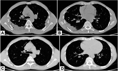
Figure 3 Biopsies: (A) transbronchial biopsies sampled several good-quality lung fragments (hematoxylin-eosin, 20× magnification). (B) A subacute lung injury was present, consisting of foci of organizing pneumonia with pneumocyte hyperplasia and several interstitial and intra-alveolar eosinophils (hematoxylin-eosin, 400× magnification). (C) In some areas, intra-alveolar foamy macrophages were intermingled with eosinophils (hematoxylin-eosin, 400× magnification). There were no extravascular granulomas, small and medium-sized vessels vasculitis.
Figure 4 Case report timeline.