
Figures & data
Table 1 Clinical Characteristics of the MRKH Patients in Two Groups
Table 2 Perioperative Data Related to Vaginoplasty in Mayer-Rokitansky-Küster-Hauser Syndrome Patients Using SIS Graft or Skin Graft
Figure 1 The surgical procedure of vaginal reconstruction for patients with MRKHS using SIS grafts. (a) A piece of swine small intestinal submucosa biological graft. (b) Shape the SIS graft into a cylinder. (c) Make incision at the mucous membrane between the urethra orifice and posterior perineum. (d) Divide the tissue between urethra and rectum to make a neovagina cavity. (e) Expand the neovagina cavity to 10cm in depth and 3 fingers in width. (f) Fix the SIS graft to neovagina cavity. (g) Place mould after operation.
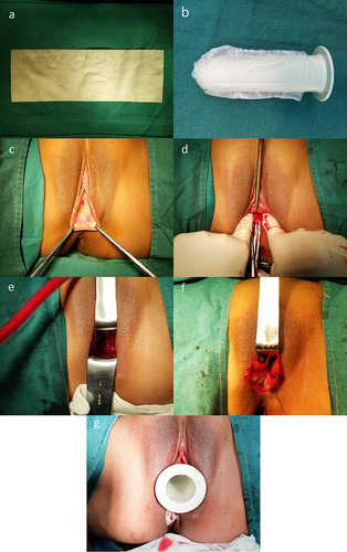
Figure 2 The surgical procedure of vaginal reconstruction for patients with MRKHS using skin graft. (a) Skin graft harvest. (b) Shape the skin graft into a cylinder.
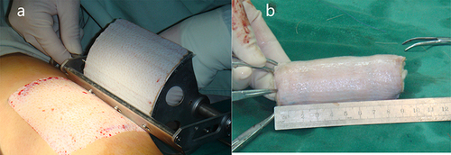
Figure 3 The colposcopy images of the neovaginal mucosa regenerative process of a patient after vaginal reconstruction using SIS graft. (a) A month later. (b) Three months later. (c) Six months later. (d) Nine months later. (e) Colpolypus of another patient.
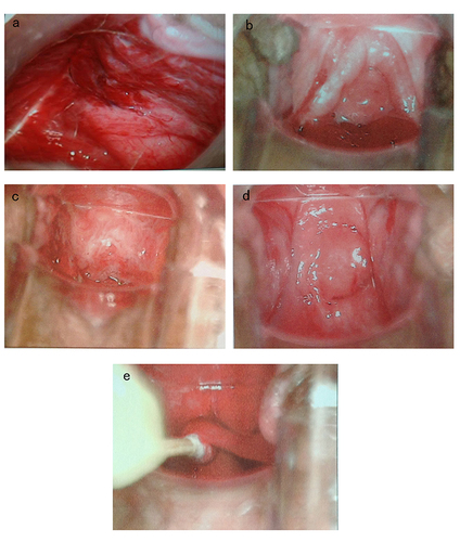
Table 3 Comparison of the Sexual Functional Outcomes in the Two Groups
Table 4 Comparison of the Anatomical Outcomes of the Two Groups After Surgery (6 Months After Surgery)