Figures & data
Figure 1 A CT pulmonary angiography (CTPA) revealed a filling defect in the pulmonary arterial phase in the right main pulmonary artery cava. and right pleural effusion.
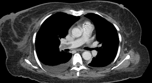
Figure 2 Contrast-enhanced CT image shows linear hypodense thrombus in IVC and bilateral renal veins.
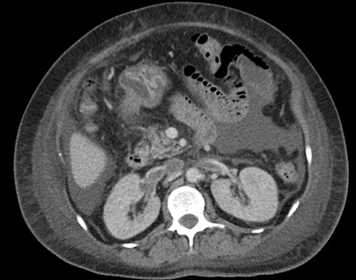
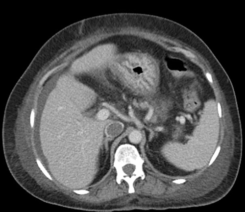
Figure 3 Contrast-enhanced CT image at the level of the diaphragm shows a filling defect (thrombus) in the inferior vena cava. and perihepatic free fluid.