Figures & data
Figure 1 Extreme brachycephalic morphology in a Pug.
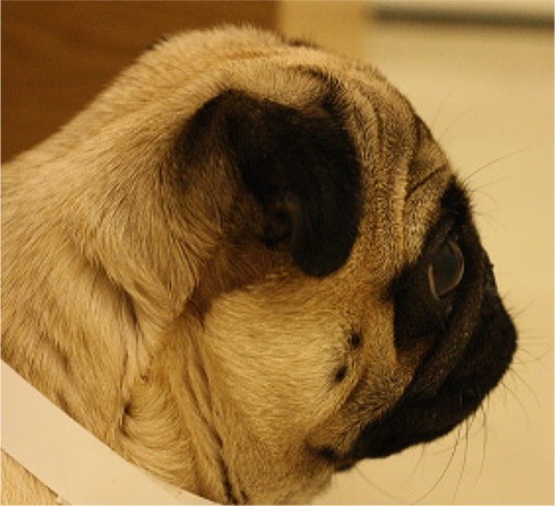
Figure 2 Pug diagnosed with brachycephalic obstructive airway syndrome exhibiting respiratory distress preoperatively.
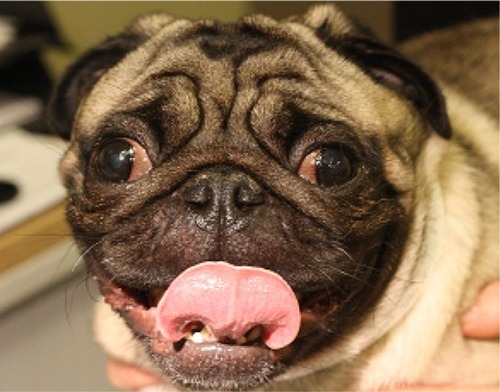
Figure 3 Elongated soft palate in a Bulldog.
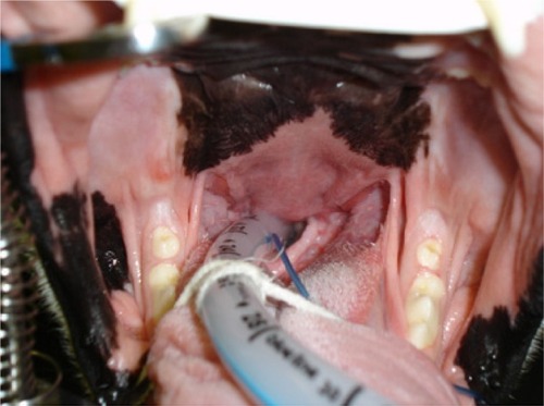
Figure 4 Stenotic nares in brachycephalic dogs, increasing in severity from left to right.
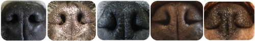
Figure 5 Close-up image of grade II laryngeal collapse.
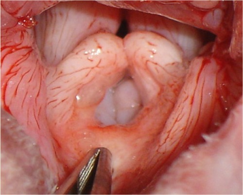
Figure 6 Lateral thoracic radiograph of a Bulldog with a hypoplastic trachea.
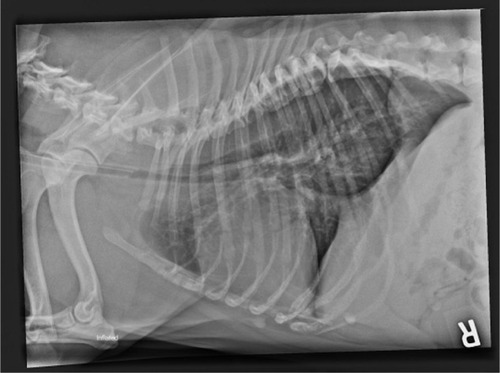
Figure 7 Elongated soft palate.
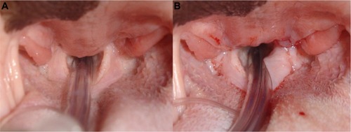
Figure 8 Stenotic nares.
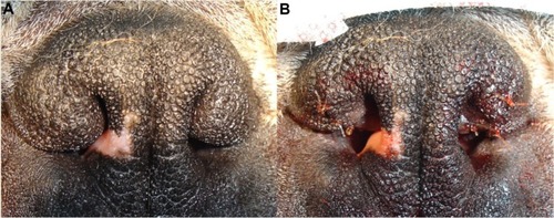
Table 1 Degree of improvement observed postsurgery in four studies of brachycephalic obstructive airway syndrome, with categories as defined by the authors of the studies divided into four outcomes
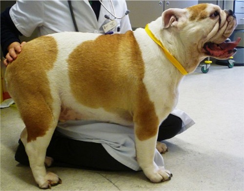
Figure 9 Obese Bulldog diagnosed with brachycephalic obstructive airway syndrome.