5,748
Views
0
CrossRef citations to date
0
Altmetric
Review

The Roles of Jumonji-Type Oxygenases in Human Disease

, , , , , & show all
Pages 89-120 | Published online: 28 Feb 2014

Figures & data

Figure 1.  Phylogenetic tree of the human 2-oxoglutarate-dependent oxygenases.

Different subfamilies discussed in the text are highlighted in various colors. Red asterisks indicate members for which no enzymatic activity has been determined yet.

Figure 1.  Phylogenetic tree of the human 2-oxoglutarate-dependent oxygenases.Different subfamilies discussed in the text are highlighted in various colors. Red asterisks indicate members for which no enzymatic activity has been determined yet.
Figure 2.  The overall fold of the catalytic JmjC domain in iron- and 2-oxoglutarate-dependent histone demethylases and nucleotide hydroxylases.

(A) Jmj prototype member JmjD2A (PDB ID: 2OQ7) in complex with Ni2+ (which replaces the endogenous Fe2+) and the 2-oxoglutarate competitive inhibitor N-oxalyl glycine (NOG). The double-stranded β-helical core elements are labeled I–VIII and colored cyan, the additional β-strands in blue and the helices in red. Ni2+ is shown as a green sphere and NOG as yellow sticks. (B) Overlay of the catalytic core (displayed are the active site metal, the Glu-His triad of active site residues and NOG) of human JmjD2A (green) compared to human ALKBH2 (PDB ID: 3BTX; light blue), indicating similar folding patterns of the catalytic domain. (C) Catalytic core of human methyladenosine demethylase FTO (PDB ID: 3LFM [Citation14]) demonstrating the double-stranded β-helical fold and including the active site metal (blue sphere).

Figure 2.  The overall fold of the catalytic JmjC domain in iron- and 2-oxoglutarate-dependent histone demethylases and nucleotide hydroxylases. (A) Jmj prototype member JmjD2A (PDB ID: 2OQ7) in complex with Ni2+ (which replaces the endogenous Fe2+) and the 2-oxoglutarate competitive inhibitor N-oxalyl glycine (NOG). The double-stranded β-helical core elements are labeled I–VIII and colored cyan, the additional β-strands in blue and the helices in red. Ni2+ is shown as a green sphere and NOG as yellow sticks. (B) Overlay of the catalytic core (displayed are the active site metal, the Glu-His triad of active site residues and NOG) of human JmjD2A (green) compared to human ALKBH2 (PDB ID: 3BTX; light blue), indicating similar folding patterns of the catalytic domain. (C) Catalytic core of human methyladenosine demethylase FTO (PDB ID: 3LFM [Citation14]) demonstrating the double-stranded β-helical fold and including the active site metal (blue sphere).

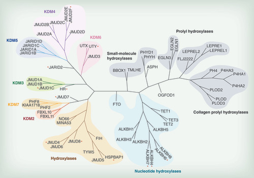
Table 1.  Domain organization and substrates of human Jmj-type oxygenases.

Table 2.   Involvement of Jmj-type demethylases and other oxygenases in human physiology and disease.