Figures & data
Created with Biorender.com.
![Figure 2. Difference in cardiac pathology between hypertrophic cardiomyopathy and dilated cardiomyopathy.Created with Biorender.com.](/cms/asset/39980537-0c70-4e6c-8367-c9adda2cc8b4/ifca_a_12326039_f0002.jpg)
Left: two-chamber delayed gadolinium enhancement imaging showing apical hypertrophic cardiomyopathy with diffuse mid-wall fibrosis in the hypertrophied segments. Right: Four-chamber SSFP imaging showing the typical appearance of dilated cardiomyopathy-related ventricular thinning.
![Figure 3. Hypertrophic versus dilated cardiomyopathy.Left: two-chamber delayed gadolinium enhancement imaging showing apical hypertrophic cardiomyopathy with diffuse mid-wall fibrosis in the hypertrophied segments. Right: Four-chamber SSFP imaging showing the typical appearance of dilated cardiomyopathy-related ventricular thinning.](/cms/asset/8a8541da-9ab8-4d9e-9d62-4ebc3db1b701/ifca_a_12326039_f0003.jpg)
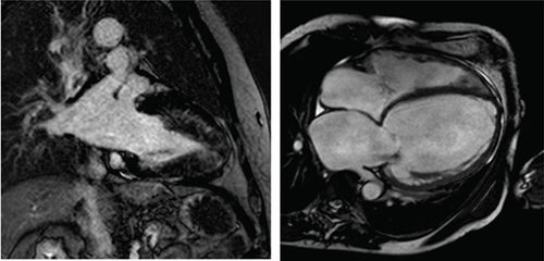
Table 1. Adapted table illustrating the results from Halliday and colleagues showing the respective hazard ratios for risk of all-cause mortality, and a composite of sudden cardiac death and aborted sudden cardiac death, for extent, pattern and location of late gadolinium enhancement.
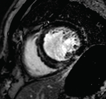
Table 2. Summary of the various roles of cardiac MRI in dilated cardiomyopathy.
![Figure 5. An illustration of ventricular wall thickening and areas of fibrosis in a heart with hypertrophic cardiomyopathy.](/cms/asset/c769b61c-fe79-4f8a-9512-629fb17d1daa/ifca_a_12326039_f0005.jpg)