
Figures & data
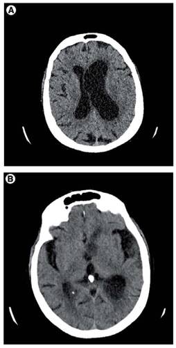
(A) Showing prominence of the ventricles with marked asymmetrical dilatation of the left lateral ventricle. (B) Layering of debris within the occipital horns.
(A) Showing prominence of the ventricles with marked asymmetrical dilatation of the left lateral ventricle. (B) Layering of debris within the occipital horns.