Figures & data
Figure 1. Bilateral subdural hematoma and diffuse pachymeningeal enhancement.
T1-weighted MRI images with gadolinium enhancement displayed the presence of bilateral subdural hematoma and widespread pachymeningeal enhancement, as observed in both coronal (A) and axial (B) sections. Additionally, axial sections of Flair MRI exhibited bilateral subdural hematoma (C).
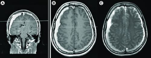
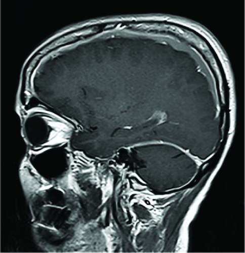
Figure 2. In the sagittal section of T1-weighted MRI with gadolinium enhancement showed diffuse pachymeningeal enhancement of the dura mater in the posterior fossa surrounding the cerebellum.