Figures & data
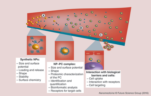
(A) The bare NP (synthetic identity) that has a specific shape, size and charge is injected into the body. (B) Once exposed to biological fluids, synthetic NPs come into contact with active biomolecules that surround them thus giving rise to the NP–PC complex (biological identity). (C) These NP–PC complexes are responsible for the interaction with biological barriers and cells. The NP can reach the target site using a specific receptor endowed upon them by the PC as part of their new biological identity (physiological response). The experimental steps associated to the study of bare NPs, NP–PC complexes and their physiological behaviors are also reported.
NP: Nanoparticle; PC: Protein corona.
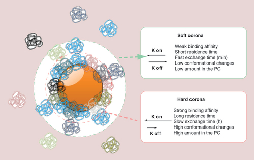
The protein corona represents the biological entity of a nanoparticle. Hard corona proteins are directly adsorbed on the nanoparticle surface due to their strong binding affinity. These proteins also have a slow exchange time. Soft corona proteins associate with the hard corona via weak protein–protein interactions, thus showing a short residence time around the nanoparticle and a fast exchange time. The objects are not drawn in scale.
PC: Protein corona.
During the very early phase: a highly complex corona is established (new). A corona of low complexity evolves slowly (old). During the late phase: the protein corona composition remains stable and shows quantitative rather than qualitative changes and binding kinetics dependent and independent of the Vroman effect (new). A highly dynamic protein corona changes significantly over time, controlled by the Vroman effect (old). The objects are not drawn to scale.
Reproduced with permission from [Citation37].
![Figure 3. Formation of the protein corona: the old versus the new model.During the very early phase: a highly complex corona is established (new). A corona of low complexity evolves slowly (old). During the late phase: the protein corona composition remains stable and shows quantitative rather than qualitative changes and binding kinetics dependent and independent of the Vroman effect (new). A highly dynamic protein corona changes significantly over time, controlled by the Vroman effect (old). The objects are not drawn to scale.Reproduced with permission from [Citation37].](/cms/asset/378d22fa-0a3f-4914-bd52-e160cf8074bc/innm_a_12340785_f0003.jpg)
The PC hides the functionalization molecule (in this case, transferrin) on the NP surface, thereby inhibiting the interaction with its specific receptor on target cells.
NP: Nanoparticle; PC: Protein corona; Tf: Transferrin.
Adapted with permission from [Citation103].
![Figure 4. Negative impact of the protein corona on the nanoparticle-targeting ability.The PC hides the functionalization molecule (in this case, transferrin) on the NP surface, thereby inhibiting the interaction with its specific receptor on target cells.NP: Nanoparticle; PC: Protein corona; Tf: Transferrin.Adapted with permission from [Citation103].](/cms/asset/23002840-2998-4b45-a0bb-48eaa103c0f1/innm_a_12340785_f0004.jpg)
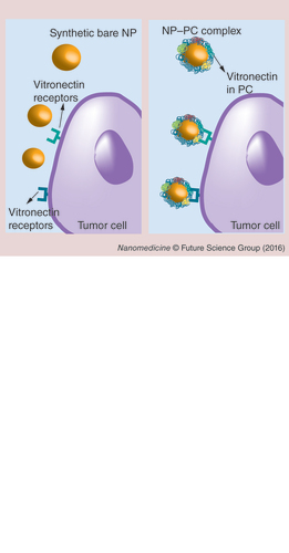
The PC contains proteins (in this case, vitronectin) that act as ligands for receptors on specific cells (tumor cells). This phenomenon, in theory, paves the way for induction of the PC formation to target selected cells. Elements not drawn to scale.
NP: Nanoparticle; PC: Protein corona.
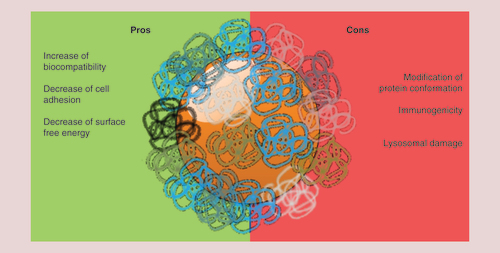
Schematic representation of the pros and cons of the protein corona in terms of cytotoxicity.
The protein corona significantly affects nanoparticle–cell interactions (e.g., cell internalization and pathway activation). The different role of protein corona in nanoparticle uptake by monocytes and macrophages is represented.
Reproduced with permission from [Citation134].
![Figure 7. Mechanisms involved in nanoparticle-induced immunomodulation.The protein corona significantly affects nanoparticle–cell interactions (e.g., cell internalization and pathway activation). The different role of protein corona in nanoparticle uptake by monocytes and macrophages is represented.Reproduced with permission from [Citation134].](/cms/asset/883be6b9-4ab1-4ada-bf11-9a382b0f2e64/innm_a_12340785_f0007.jpg)
The transfer of cellular membrane properties to synthetic NPs led to the development of the so-called biomimetic strategies. Specialized cell types (A), such as red blood cells [Citation155], leukocytes [Citation156] or platelets [Citation157], inspired the development of particles that share a similar cellular membrane composition. The resulting vectors, defined as ‘bio-inspired NPs’, are recognized as self by the immune system. This could depend on the protein corona (PC) adsorbed on their surface. To date, no studies have been performed in order to describe the PC of bio-inspired NPs. (B) We hypothesize that while a synthetic NP is mainly recognized as nonself and adsorbs many serum proteins; bio-inspired NPs (reported to possess cell-like capabilities [Citation155–157]) absorb a specific PC similar to the PC of the cells used as membrane source for the NP synthesis. This confers self-like properties to the bio-inspired NPs and mediates their biological activity.
NP: Nanoparticle.
![Figure 8. Protein corona of bio-inspired nanoparticles.The transfer of cellular membrane properties to synthetic NPs led to the development of the so-called biomimetic strategies. Specialized cell types (A), such as red blood cells [Citation155], leukocytes [Citation156] or platelets [Citation157], inspired the development of particles that share a similar cellular membrane composition. The resulting vectors, defined as ‘bio-inspired NPs’, are recognized as self by the immune system. This could depend on the protein corona (PC) adsorbed on their surface. To date, no studies have been performed in order to describe the PC of bio-inspired NPs. (B) We hypothesize that while a synthetic NP is mainly recognized as nonself and adsorbs many serum proteins; bio-inspired NPs (reported to possess cell-like capabilities [Citation155–157]) absorb a specific PC similar to the PC of the cells used as membrane source for the NP synthesis. This confers self-like properties to the bio-inspired NPs and mediates their biological activity.NP: Nanoparticle.](/cms/asset/aa3ea1da-7dc1-4e77-9400-52003931fd06/innm_a_12340785_f0008.jpg)
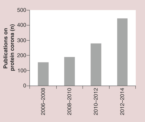
The number of articles has tripled during the 3 years between 2012–2014 versus 2006–2008.