
Figures & data
Figure 1. Pt #1: Male 48 years of age. Endoscopic view of the gastric tumour located in the fundus.
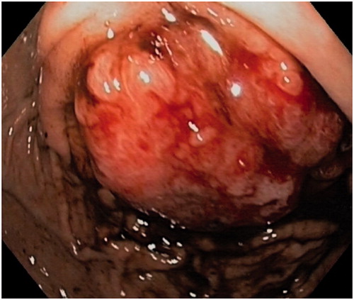
Figure 2. Time trends of the AFP levels in Pt #1 during the trajectory of his disease.
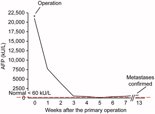
Figure 3. Pt #1: Coronal multi detector computer tomography(MDCT) shows the gastric tumour (T) in the left hypochondrium with infiltration of the left hemidiaphragma (stapled arrow) and the greater curvature (arrow) of the stomach.
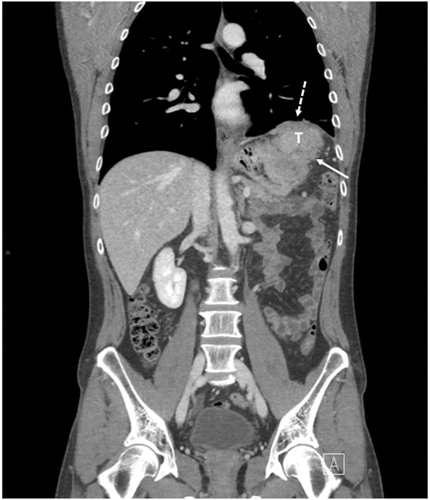
Figure 4. (A) Histopathology of hepatoid carcinoma in the gastric mucosal primary lesion The tumour comprised a mixture of pseudoglandular and hepatoid components. Note the normal glandular structure (arrows) (haematoxylin and eosin staining; original magnification, ×400). (B) Immunohistochemical staining showing intracytoplasmic positivity for AFP (dark brown). Note the non-staining normal glandular structure (arrows) (original magnification, x400).
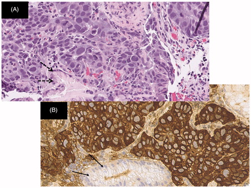
Figure 5. Pt # 2: Female 81 years of age. (A) Axial MDCT at the time of diagnosis showing the gastric tumour (T) located in the antrum, with lymph nodes (LN) at the lesser curvature. (B) Five months after primary diagnosis: disease progression with liver metastasis (M), tumour and lymph node growth (T) along the lesser curvature, ascites (A) and thrombus in portal veins (white arrow).
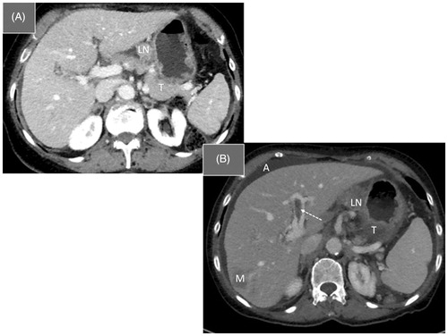
Table 1. Patient characteristics and outcomes presented in five recent series.