Figures & data
Figure 1. Chemical structure and sites of radiolabel for [14C]-peginesatide.
![Figure 1. Chemical structure and sites of radiolabel for [14C]-peginesatide.](/cms/asset/9e58bd9d-4fd3-4f39-9f34-fd7658c97ec2/ixen_a_649310_f0001_b.gif)
Table 1. Mean pharmacokinetic parameters of peginesatide in the plasma of male Sprague–Dawley rats after IV administration.
Figure 2. The plasma concentration–time profiles of peginesatide in male Sprague–Dawley rats after a single IV administration of peginesatide at a dose of 0.1, 0.5, or 5 mg·kg−1. Plasma concentration of peginesatide was determined by ELISA. Each data point represents the mean ± SD for three rats.
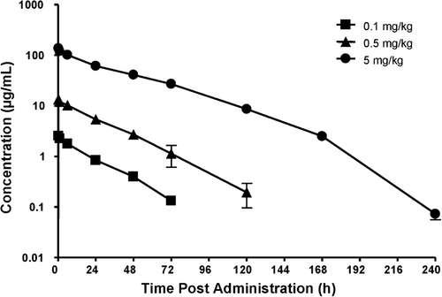
Table 2. Concentrations of the radioactivity in the tissues of albino rats after a single IV administration of 5 mg·kg−1 [14C]-peginesatide.
Figure 3. Log plasma concentration of the peginesatide-derived radioactivity in the plasma, eyes and skin of male albino (Sprague–Dawley) and pigmented (Long Evans) rats as a function of time after IV administration of 5 mg·kg−1 [14C]-peginesatide. Each value represents the mean ± SD for three animals.
![Figure 3. Log plasma concentration of the peginesatide-derived radioactivity in the plasma, eyes and skin of male albino (Sprague–Dawley) and pigmented (Long Evans) rats as a function of time after IV administration of 5 mg·kg−1 [14C]-peginesatide. Each value represents the mean ± SD for three animals.](/cms/asset/d110c973-46fb-49de-95d8-9d1e5f0b27c2/ixen_a_649310_f0003_b.gif)
Figure 4. Whole-body sections and subsequent autoradiographs of male Sprague–Dawley rats 72 h, 2 and 4 weeks following IV administration of 17 mg·kg−1 [14C]-peginesatide. Dark areas represent high radioactive concentrations.
![Figure 4. Whole-body sections and subsequent autoradiographs of male Sprague–Dawley rats 72 h, 2 and 4 weeks following IV administration of 17 mg·kg−1 [14C]-peginesatide. Dark areas represent high radioactive concentrations.](/cms/asset/2d8e89e6-1b22-4a68-add5-8fd602bea6ed/ixen_a_649310_f0004_b.gif)
Figure 5. Biolocalization of peginesatide to hematopoietic and EMH sites compared to muscle following a single IV administration of 17 mg·kg−1 [14C]-peginesatide. Each data point represents the mean ± SD for three rats.
![Figure 5. Biolocalization of peginesatide to hematopoietic and EMH sites compared to muscle following a single IV administration of 17 mg·kg−1 [14C]-peginesatide. Each data point represents the mean ± SD for three rats.](/cms/asset/57c1f231-f3fa-46a0-a2da-512bdf9c8ded/ixen_a_649310_f0005_b.gif)
Figure 6. Cumulative recovery of peginesatide-related radioactivity in urine and feces of male rats following IV administration of 5 mg·kg−1 [14C]-peginesatide. At 2 weeks after dosing, the remaining approximately 50% of the radioactivity not excreted was detected in the carcass. Each value represents the mean ± SD for three animals.
![Figure 6. Cumulative recovery of peginesatide-related radioactivity in urine and feces of male rats following IV administration of 5 mg·kg−1 [14C]-peginesatide. At 2 weeks after dosing, the remaining approximately 50% of the radioactivity not excreted was detected in the carcass. Each value represents the mean ± SD for three animals.](/cms/asset/c8061f36-c3bb-4b8e-aede-e8d9389b3d68/ixen_a_649310_f0006_b.gif)
Table 3. Pharmacokinetic parameters of peginesatide and associated radioactivity in plasma after single IV administration of [14C]-peginesatide at a dose of 5 mg·kg−1 to rats.
Table 4. Percent dose excretion of [14C]-peginesatide and associated radioactivity in rat urine and feces after single IV administration of 5 mg·kg−1.
Table 5. Day 14 concentrations of peginesatide and associated radioactive compounds in rat plasma and tissues after a single IV administration of 5 mg/kg [14C]-peginesatide.