Figures & data
Figure 1. Geometry of the proposed antenna. (A) Cross-sectional top view. (B) Cross-sectional side view.
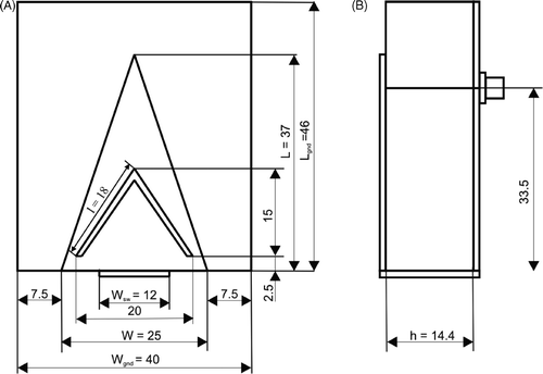
Figure 2. Visualisation of the single antenna setup.
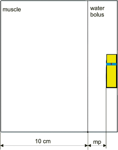
Figure 3. Visualisation of the 12-antenna set-up.
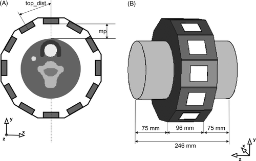
Table I. Dielectric parameters of Cole–Cole dispersion model used to predict the dielectric properties in the selected neck structures.
Figure 4. Simulated return loss for various permittivities of matching liquid.
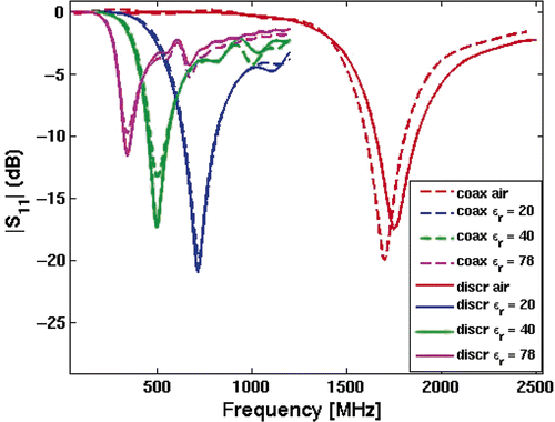
Figure 5. E-field distribution along the patch at different frequencies. Cross-sectional top view. (A) 350 MHz; (B) 800 Mhz.
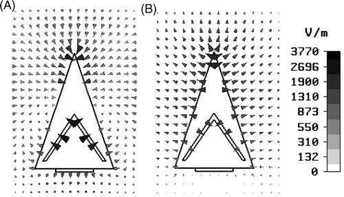
Figure 6. SAR distribution in muscle phantom for different frequencies. (A) f = 350 MHz; (B) f = 800 MHz.
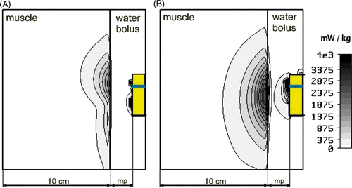
Figure 7. Simulated return loss for various distances between the muscle and the antenna.
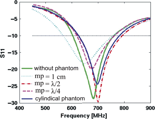
Figure 8. Simulated return loss for different couplings between antennas.
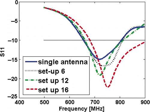
Table II. The areas (mm2) enclosed by centrally located 50%, 75% and 90% iso-SAR contours and the length and width of the focus for set-ups of 12 and 16 antennas at various frequencies.
Figure 9. The normalised SAR distributions calculated in the cross-section through the muscle phantom at y = 0 cm for (A) 12-antenna set-up, f = 500 MHz; (B) 12-antenna set-up, f = 800 MHz; (C) 16-antenna set-up, f = 500 MHz; (D) 16-antenna set-up, f = 800 Mhz. The axes are in mm.
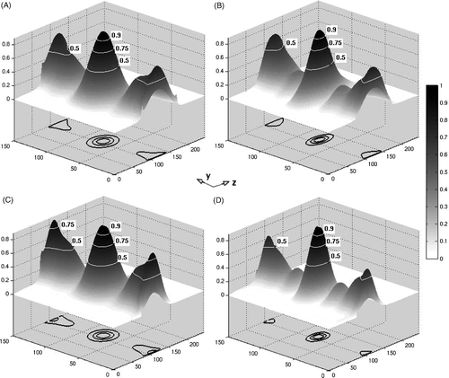
Figure 10. The normalised SAR distributions calculated in the cross-section through the semi-3D neck phantom containing tumour at z = 110 mm. The axes are in mm. (A)12-antenna set-up, frequency f = 500 MHz; (B)12-antenna set-up, f = 800 MHz; (C)16-antenna set-up, d = 16.5 cm, f = 500 MHz; (D)16-antenna set-up, d = 16.5 cm, f = 800 Mhz. (E)16-antenna set-up, d = 14 cm, f = 500 MHz; (F) 16-antenna set-up, d = 14 cm f = 800 Mhz.
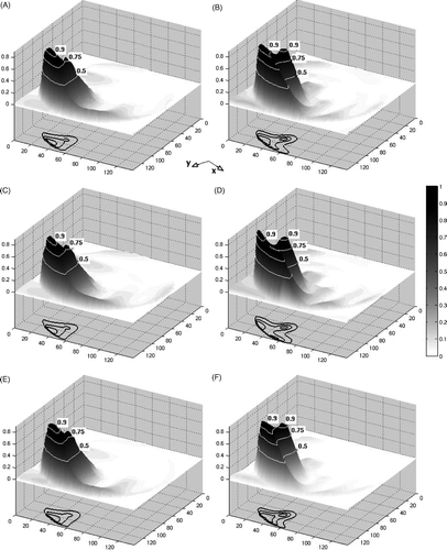
Table III. aPA ratio calculated in 2D and 3D for phantom both with and without tumour. Frequency f = 800 Mhz.
Table IV. The areas (mm2) enclosed by 75% and 90% iso-SAR contours in the target for set-ups of 12 and 16 antennas at various frequencies.
Figure 11. The average power absorption (aPA) ratio as a function of frequency for 12- and 16-antenna arrangements, calculated for both 2D and 3D.
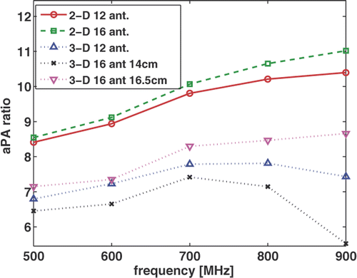
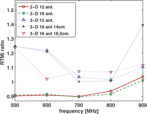
Figure 12. The remaining tissue maximum index (RTMi) as a function of frequency for 12- and 16-antenna arrangements, calculated in both 2D and 3D.