Figures & data
Figure 1. Hsp27 and Hsp70 expression measured 16 h following laser irradiation for three tumour depths with distinct temperature elevation zones (top left) and corresponding mean MRTI measured maximum temperature elevation during laser irradiation of five mice (top right). Mean measured temperature elevation as a function of time at the tumour surface (bottom centre).
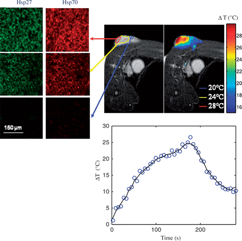
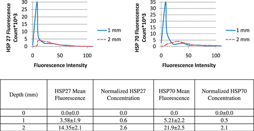
Figure 2. Histograms for quantification of Hsp27 and Hsp70 fluorescence and corresponding Hsp27 and Hsp70 mean fluorescence, standard deviation, and normalised concentration at tumour depths of 0, 1, 2 mm from the tumour surface measured 16 h following laser irradiation.
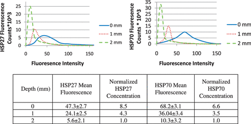
Figure 3. Fluorescence images of Hsp27 before (A) and following nanoshell inclusion (B), Hsp70 before (C) and following nanoshell inclusion (D) and histogram of Hsp27 and Hsp70 fluorescence counts as a function of fluorescence intensity following nanoshell inclusion (E).
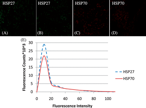
Figure 4. Hsp27 and Hsp70 expression dual stained image for three distinct tumour depths (top left) measured 16 h following laser irradiation in combination with nanoshells and corresponding MRTI measured mean maximum temperature increase for five mice (top right). Average change in temperature versus time at the tumour surface (bottom centre).
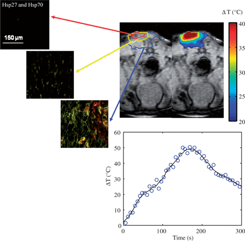
Figure 5. Histograms for quantification of Hsp27 and Hsp70 fluorescence and corresponding Hsp27 and Hsp70 mean fluorescence, standard deviation, and normalised concentration at tumour depths of 0, 1, 2 mm measured 16 h following laser irradiation with nanoshells.