Figures & data
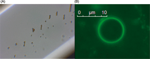
Figure 1. Theoretical structure of a magnetic microbubble based upon the formulation by Stride et al. The gas core is surrounded by a ferrofluid and stabilized by an outer coating of L-α-phosphatidylcholine. The average size is about 2 µm.
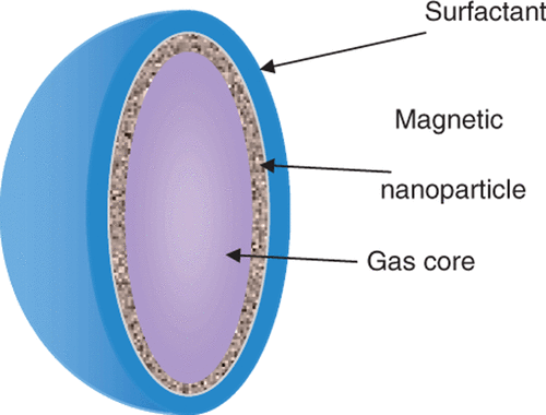
Figure 2. Bioluminescence imaging data for transfection enhancement of Chinese hamster ovary cells by magnetic microbubbles under different exposure conditions (101). A = non-magnetic microbubbles; B = magnetic micelles; C(i) = magnetic microbubbles. US = ultrasound; MF = magnetic field. Up/down refers to the surface on of the exposure chamber on which the cells were located and hence whether cell/bubble contact would be promoted by buoyancy.
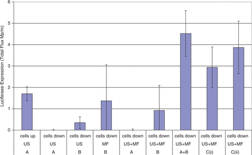
Figure 3. (A) Phase contrast and (B) fluorescence (measured at a wavelength of 490/509 nm) microscopy images of the Magnetic and acoustically active liposomes composed of fluidMAG-Tween-60 magnetic nanoparticles and the luciferase plasmid pBLuc, which was fluorescently labelled with the intercalated dye YOYO-1. Reproduced with kind permission from John Wiley & Sons from of reference Citation[106] © 2010 WILEY-VDH Verlag GmbH & Co. KGaA, Weinheim.
![Figure 3. (A) Phase contrast and (B) fluorescence (measured at a wavelength of 490/509 nm) microscopy images of the Magnetic and acoustically active liposomes composed of fluidMAG-Tween-60 magnetic nanoparticles and the luciferase plasmid pBLuc, which was fluorescently labelled with the intercalated dye YOYO-1. Reproduced with kind permission from John Wiley & Sons from Figure 2 of reference Citation[106] © 2010 WILEY-VDH Verlag GmbH & Co. KGaA, Weinheim.](/cms/asset/56a3cc63-5800-4d1f-a279-6561c4cae42f/ihyt_a_668639_f0003_b.gif)
Figure 4. Magnetic microbubbles prepared by mixing non-magnetic microbubbles with fluorescently labelled magnetic micelles. (A) retention against flow in a capillary tube following application of a magnetic field (B) fluorescent microbubble obtained by mixing non-fluorescent microbubbles with fluorescent magnetic micelles.