Figures & data
Table 1. The mitochondrial antioxidant system.
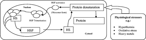
Figure 1. Generation and targets of reactive oxygen species in mitochondria. (a) A schematic diagram of ROS generation and their targets under thermal neutral conditions. Solid arrows indicate generation and diffusion of molecules. Dashed arrows indicate attenuated/blocked reactions. (b) An illustration of ROS generation and their targets under heat stress conditions. GSH depletion leads to increasing and OH√ production, and therefore mitochondrial damage.
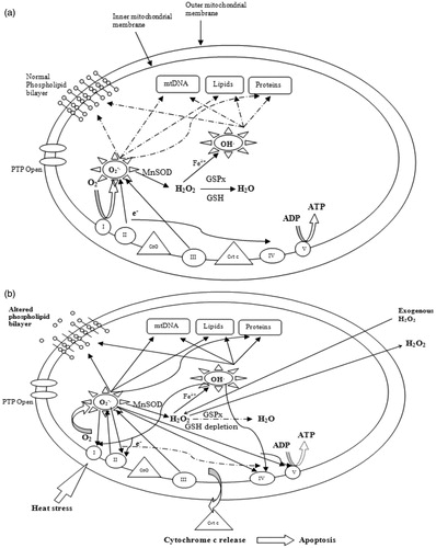
Figure 2. A schematic diagram of the different steps of the peroxidation of PUFA. This reaction is initiated when ROS abstract a hydrogen atom from the PUFA, leading to the generation of a peroxyl radical and the production of lipid free radicals.
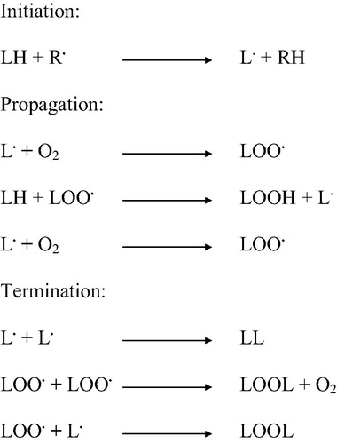
Figure 3. A schematic diagram of the mechanism of HSP synthesis. Denatured proteins accumulate in response to physiological stressors, and activate HSFs. HSFs are hyperphosphorylated in the nucleus and bind to the HSEs. HSP mRNA is then transcribed. HSP are synthesized and ensure the renaturation of denatured proteins.