Figures & data
Table 1. Effect of DHA administration on body weight, serum total protein, serum urea, serum TAG, serum TC, urinary protein, urinary NAG, and Ccr modified by CsA treatment in rats
Figure 1. Effect of DHA on the level of TBARs (index of lipid peroxidation) in the kidney of control, DHA, and CsA nephrotoxicity groups. Data are presented as mean ± SD, n = 12. Multiple comparisons were achieved using one-way ANOVA followed by Tukey–Kramer as post-ANOVA test.
Note: a and b indicate significant change from control, DHA, and CsA nephrotoxicity groups, respectively, at p < 0.05.
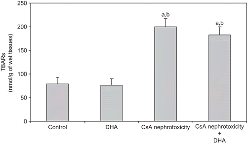
Figure 2. Effect of DHA on the level of PCC (index of protein oxidation) in the kidney of control, DHA, and CsA nephrotoxicity groups. Data are presented as mean ± SD, n = 12. Multiple comparisons were achieved using one-way ANOVA followed by Tukey–Kramer as post-ANOVA test.
Note: a and b indicate significant change from control, DHA, and CsA nephrotoxicity groups, respectively, at p < 0.05.
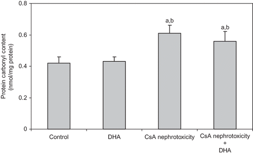
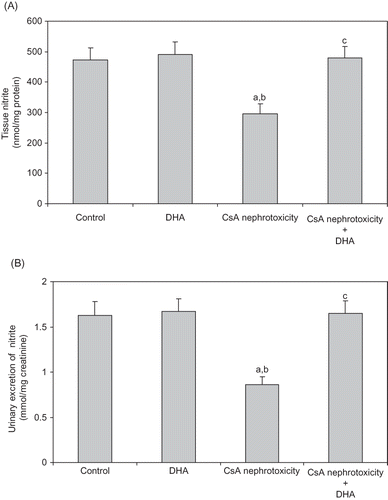
Figure 3. Effect of DHA administration on the renal tissue nitrite (A), and urinary excretion of nitrite (B) modified by CsA treatment in rats. Data are presented as mean ± SD, n = 12. Multiple comparisons were achieved using one-way ANOVA followed by Tukey–Kramer as post-ANOVA test.
Note: a, b, and c indicate significant change from control, DHA, and CsA nephrotoxicity groups, respectively, at p < 0.05.