Figures & data
Table 1. Primer sequences for PCR assays in this study.
Figure 1. The Blimp-1 mRNA expression in PMBCs of mice at 3 weeks after administration of the lentivirus Blimp-1 siRNA (study group) or PLL3.7 (control group). PMBCs of the mice (8 mice in each group) were collected, and mRNA expression of Blimp-1 detected by RT-PCR. C: control group, S: study group.
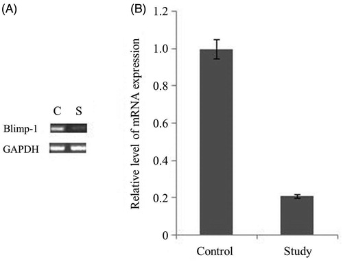
Figure 2. The expression levels of Blimp-1 protein in the kidney, liver, lymph nodes and spleen in the experimental groups. (A) 15-week-old MRL-Fas(lpr) mice received an intravenous tail vein injection of lentivirus vector. After 21 days, the mice were sacrificed, and the Blimp-1 expression in kidney, spleen, lymph node and liver was analyzed by Western blot. (B) Blimp-1 expression was analyzed by semi-quantitative Western blot by using GAPDH for normalization. *Compared with controls, p < 0.05. C: control group, S: study group. The results are representative of three individual experiments.
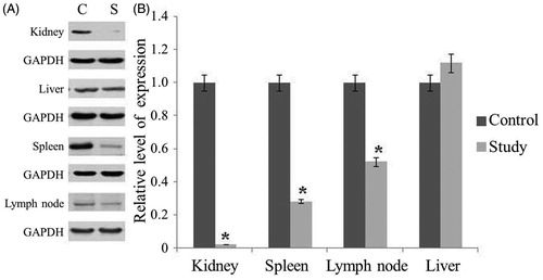
Figure 3. Immunohistochemical staining of Blimp-1. C: control group, S: study group. The numbers of Blimp-1 positive cells (marked by black arrows) in glomerulus, renal tubular epithelium, spleen, and lymph nodes of the control group were obviously greater than those in the Blimp-1 siRNA-treated group. Blimp-1 expression of blood, kidney, spleen and lymph node was decreased significantly following Blimp-1 siRNA administration.
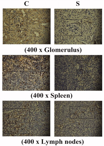
Figure 4. The level of anti-dsDNA Ab in serum of the 18-week-old MRL-Fas(lpr) mice. At 15 weeks old, MRL-Fas(lpr) mice (8 mice per group) received an intravenous tail vein injection of lentivirus (1×10⁹ pfu) or controls. The anti-dsDNA Ab concentrations in mouse serum were determined at 3-week intervals. Similar results were obtained in three individual experiments. *p < 0.05 compared with control.
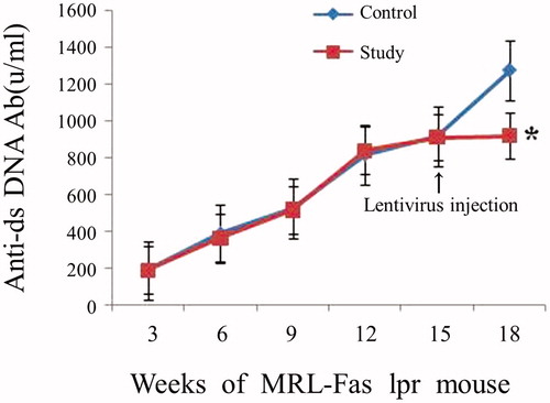
Figure 5. The effect of Blimp-1 inhibition on the expression of BCMA, XBP-1, J-chain, and C-myc. Western blot and RT-PCR were performed 3 weeks after administration of the lentivirus Blimp-1 siRNA (study group) or PLL3.7 (control group). (A) Peripheral blood of the mice (8 mice per group) was collected, and mRNA expression of Blimp-1, BCMA, XBP-1, J-chain, and C-myc were detected by RT-PCR. (B) Spleen cells of mice were collected, and protein expression of XBP-1 and C-myc were detected by Western blot. Similar results were obtained from three individual experiments. C: control group, S: study group.
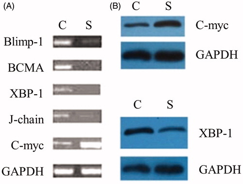
Table 2. The 24-h urinary protein concentration in experimental groups.
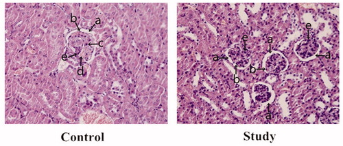
Figure 6. The renal pathological changes in the experimental groups. (a) capsular space; (b) endothelial cells; (c) lymphocytes; (d) capillary; (e) mesangial cells. In the control group, the numbers of glomerular mesangial and endothelial cells were increased compared to the study group, and glomerular capillary was dilated and congested with mild lymphocytic infiltration, which further narrowed the capsular space. After Blimp-1 expression was inhibited by Blimp-1 siRNA (study group), the glomerular pathological changes in the study group were mitigated. The numbers of mesangial and endothelial cells and the capsular space tended to be normal, and there were no significant inflammatory changes found in glomerulus.