Figures & data
Table 1. Influence of psychological stress on cardiovascular parameters in healthy male volunteers (N = 31).
Figure 1. Acute psychological stress influences T cells expressing Th1 or Th2 phenotypes. The stress-induced redistribution of CRTH2+ and CD226+ T cell subsets was analyzed in healthy male subjects (N = 31) after resting phase 1 (pre), immediately after completion of the stress test (stress), and following the final resting phase (post) using four-color flow cytometry. Analysis was performed using a combination of a morphological lymphocyte gate and gates for CD4+ T cells (CD3+CD4+) and CD8+ T cells (CD3+CD8+), respectively. Asterisks indicate significant (**p < 0.01, ***p < 0.001) differences compared to baseline values as determined by Wilcoxon’s rank sum test (A). Dot plots representative for CCR7 versus CD226 staining of CD4+ and CD8+ T cells are shown for one test subject (B).
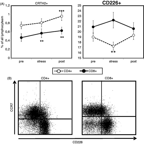
Figure 2. Acute psychological stress causes a short-lasting increase in circulating γδ T cells. Percentages of circulating T cells expressing the γδ T cell receptor (TCR) were determined in healthy male subjects (N = 31) after resting phase 1 (pre), immediately after completion of the stress test (stress), and following the final resting phase (post) using four-color flow cytometry. Analysis was performed using a combination of a morphological lymphocyte gate and gates for CD4+ T cells (CD3+CD4+) and CD8+ T cells (CD3+CD8+). Percentages of CD4 and CD8 double-negative T cells expressing the γδ TCR were then measured using an appropriate isotype control. Asterisks indicate significant (**p < 0.01) differences compared to baseline values as determined by Wilcoxon’s rank sum test.
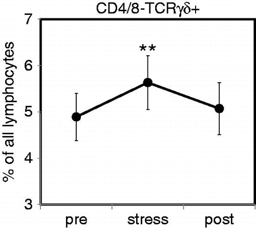
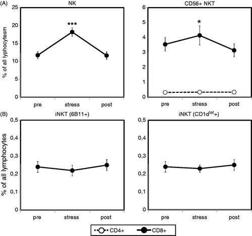
Figure 3. Peripheral numbers of conventional CD56+ NKT cells, but not invariant chain NKT (iNKT) cells, increase after acute psychological stress. Percentages of circulating NK cells, CD56+ NKT cells (A) and iNKT cells (B) were determined in healthy male subjects (N = 31) after resting phase 1 (pre), immediately after completion of the stress test (stress) and following the final resting phase (post) using four-color flow cytometry. Analysis was performed using a combination of a morphological lymphocyte gate and gates for NK cells (CD3−CD56+), conventional NKT cells (CD3+CD8+CD56+ or CD3+CD4+CD56+) and iNKT (CD3+6B11+ or CD3+CD1d-tetramer+). Percentages of the respective lymphocyte subsets were measured using appropriate isotype controls. Asterisks indicate significant (*p < 0.05, ***p < 0.001) differences compared to baseline values as determined by Wilcoxon’s rank sum test.