Figures & data
Table I. Inotropic and automatic responses to isoproterenol (ISO) and norepinephrine (NE) in ventricular myocytes from control rats (CTR) and rats subjected to restraint (RST) and footshock (FS) for 3 days.
Figure 1 Concentration-effect curves for norepinephrine (NE) in ventricular myocytes isolated from control rats (CTR; N = 27), as well as rats submitted to restraint (RST; N = 15) and footshock (FS; N = 27) sessions for 3 days. Inotropic response (panel A) was taken as the increase in peak twitch cell shortening (ΔL), expressed as percent of the resting cell length (RL). Automatic response (panel B) was taken as the increase in the rate of spontaneous contractions (SC) in the absence of electric stimulation. Symbols and bars indicate mean and SEM values, respectively. Curve parameters are presented in .
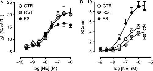
Table II. Inotropic and automatic responses in ventricular myocytes from control (CTR) and footshock-stressed rats (FS): influence of β-adrenoceptor antagonists and response to 3-isobutyl-1-methylxanthine (IBMX).
Figure 2 Concentration-effect curves for isoproterenol (ISO) in ventricular myocytes isolated from control (CTR) and footshock-stressed rats (FS), determined in the presence of 0.1 μM ICI118551 (ICI). Dashed gray and black lines indicate the curve to ISO obtained in the absence of the antagonist in CTR and FS, respectively. Twitch shortening peak (panel A) and rate of spontaneous contractions during rest (panel B) are expressed as in Figure 1. Panel C shows the rate of extrasystolic contractions (ES) recorded during pacing as a function of ISO concentration. Symbols and bars indicate mean and SEM values, respectively. Curve parameters are presented in .
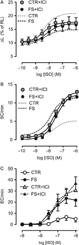
Figure 3 Inotropic (left bars: increase in twitch peak shortening, ΔL, as percent of resting cell length, RL) and automatic (right bars: rate of spontaneous contractions, SC, at rest) responses to 3 μM forskolin (FSK) in ventricular myocytes isolated from control (CTR) and footshock-stressed (FS) rats. Bars are mean and SEM. Baseline ΔL was 8.0 ± 1.1 in CTR (N = 19) and 8.9 ± 0.8% of RL in FS (N = 20). Baseline SC rate was 0.3 ± 0.2 and 0.8 ± 0.3 SC/min in CTR and FS, respectively (p>0.10). *p < 0.05 vs CTR (t-test for unpaired samples).
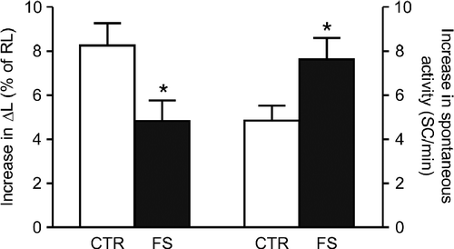
Figure 4 Concentration-effect curves for 3-isobutyl-1-methylxanthine (IBMX) in ventricular myocytes isolated from control (CTR; N = 9) and footshock-stressed rats (FS; N = 12). Inotropic (panel A) and automatic (panel B) responses are expressed as in Figure 1. Symbols and bars indicate mean and SEM values, respectively. Baseline ΔL and SC rate were not significantly different in CTR and FS (p>0.36). Curve parameters are presented in .
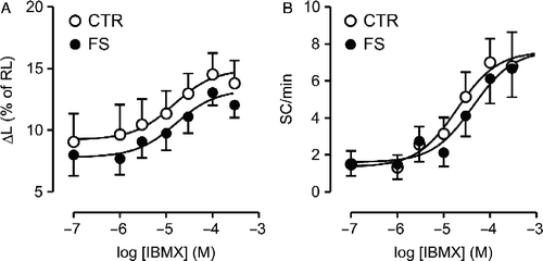
Figure 5 Inotropic (panel A) and automatic (panel B) responses to the increase in extracellular CaCl2 in isolated ventricular myocytes from control (CTR; N = 8) and footshock-stressed rats (FS; N = 8). Symbols and bars indicate mean and SEM values, respectively.
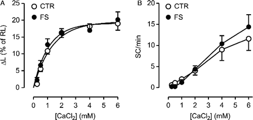
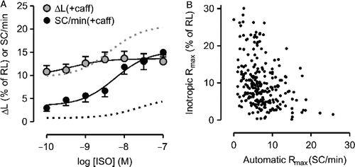
Figure 6 A: Concentration-effect curve for isoproterenol (ISO) determined in the presence of 1 mM caffeine (caff). Inotropic and automatic responses were determined in the same set of control ventricular myocytes (N = 8) and are expressed as in Figure 1. Data are mean and SEM. Gray and black dashed lines indicate the curves for the inotropic and automatic effects of ISO, respectively, in the absence of caffeine. B: Relationship between automatic and inotropic maximum responses (Rmax) to agonists determined in a set of 205 myocytes.