Figures & data
Figure 1. Structure of fluorescein isothiocyanate dextran 4000.
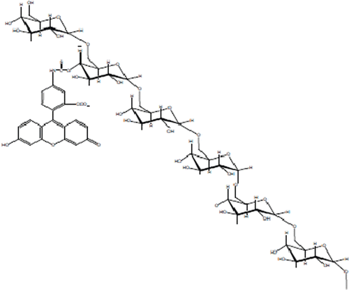
Figure 2. Microdialysis experimental design.
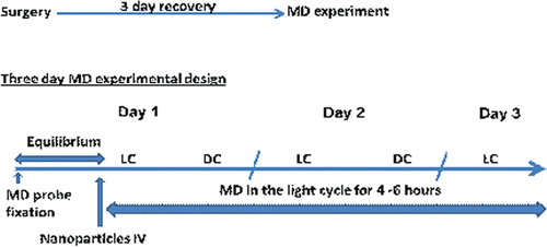
Figure 3. Transmission electron microscopic pictures of pegylated nanoparticles.
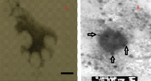
Figure 4. Distribution of FD4 in various organs after exposure to (A) FD4 saline solution, (B) Peglyated nanoparticles containing FD4, (C) Antibody-coated pegylated nanoparticles containing FD4.
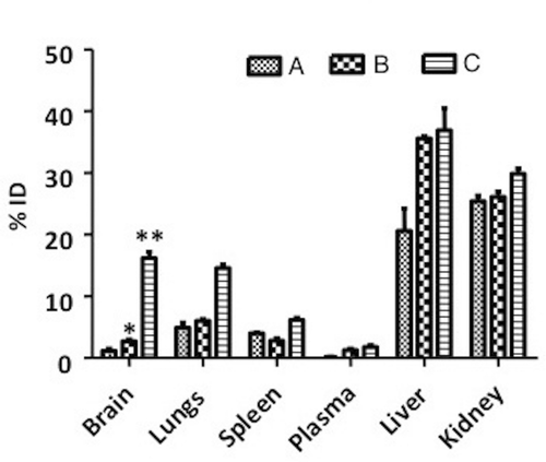
Figure 5. Three-day in vivo brain microdialysis results: concentration of FD4 (corrected for probe recovery) in VHIP of animals receiving antibody-coated pegylated nanoparticles containing FD4. VHIP, ventral hippocampus.
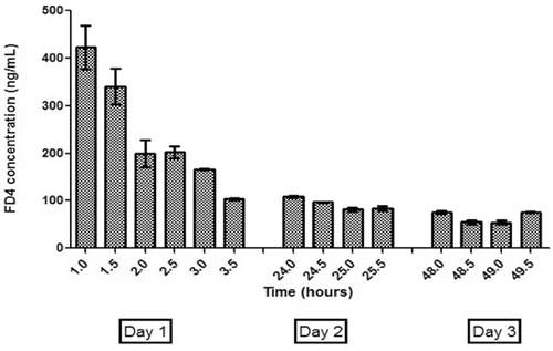
Figure 6. Immunofluorescence confocal imaging of animal tissue after exposure to (A) FD4 saline solution, (B) Peglyated nanoparticles containing FD4, (C) Antibody-coated pegylated nanoparticles containing FD4. Green: FD4, Red: Secondary antibody associated with TRITC.
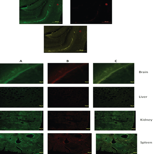
Figure 7. Fluorescence microscopic picture of animal tissue after exposure to (A) FD4 saline solution, (B) Peglyated nanoparticles containing FD4, (C) Antibody-coated pegylated nanoparticles containing FD4.
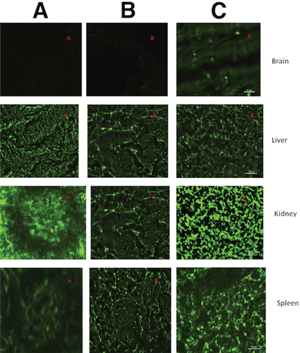
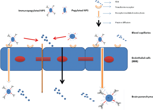
Figure 8. Possible mechanisms to explain targeting efficacy of antibody-coated pegylated nanoparticles.