Figures & data
Figure 1. Synthesis of non-modified and Oct-modified HPMA copolymer conjugates with Dox.
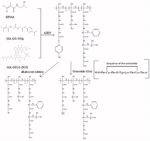
Table 1. Characteristics of the synthesized HPMA copolymers.
Figure 2. Quantitative PCR analysis of expression levels of SSTR2 in HepG2, A549 and H22 cells.
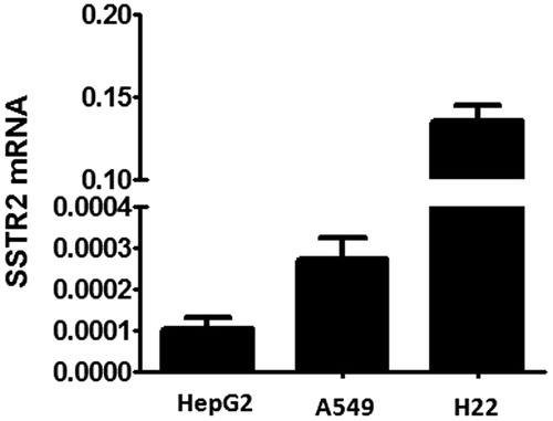
Figure 3. Internalization of P-FITC and P-FITC-Oct (equivalent to 50 mM of FITC) co-incubated with or without free Oct (0.04 mg/ml) in HepG2 (A–D) and A549 (E–H) cells by inverted fluorescence microscopy. The green signal indicated FITC-labeled conjugates while blue signal indicated nuclei. A: HepG2 cells incubated with P-FITC; B: HepG2 cells incubated with P-FITC and free Oct; C: HepG2 cells incubated with P-FITC-Oct; D: HepG2 cells incubated with P-FITC-Oct and free Oct; E: A549 cells incubated with P-FITC; F: A549 cells co-incubated with P-FITC and free Oct; G: A549 cells co-incubated with P-FITC-Oct; H: A549 cells incubated with P-FITC-Oct and free Oct.
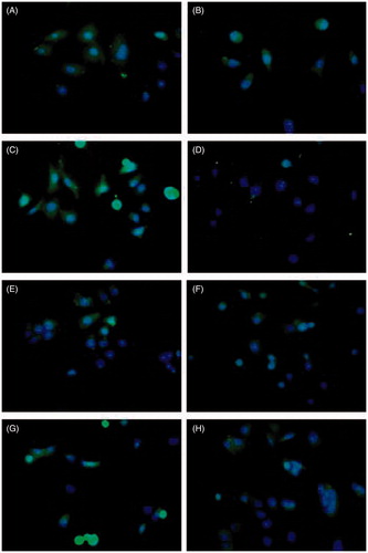
Figure 4. Quantitative study of uptake of P-DOX and P-DOX-Oct in HepG2 and A549 cells in presence or absence of free Oct. A: cells were incubated with various concentrations of conjugates for 2 h. B: cells were incubated with conjugates (0.6 mg polymer/ml) and 0.04 mg/ml of free Oct (#p < 0.05 for P-DOX-Oct versus P-DOX-Oct with free Oct).
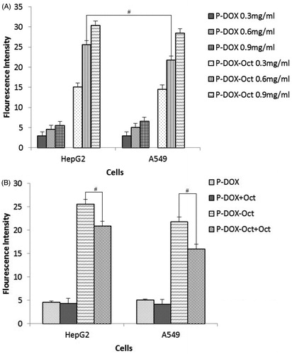
Figure 5. Morphological and nuclear changes of HepG2 (A-C) and A549 (D-F) cells treated with P-DOX and P-DOX-Oct for 24 h as determined by DAPI staining. A and D: control groups. B and E: P-DOX treated groups. C and F: P-DOX-Oct treated groups. The white arrows pointed to the apoptosis and necrosis.
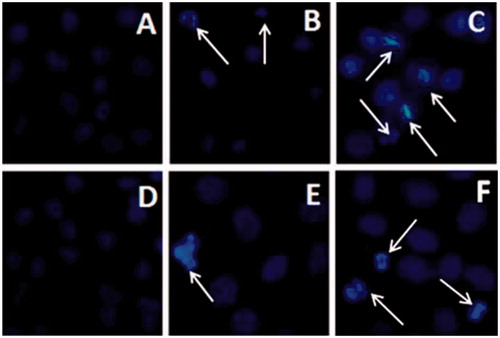
Table 2. The IC50 values of DOX, P-DOX and P-DOX-Oct against HepG2 and A549 cells (n = 3).
Figure 6. In vivo biodistribution of non-modified (A) and Oct-modified (B) HPMA copolymers at 24, 72 and 120 h.
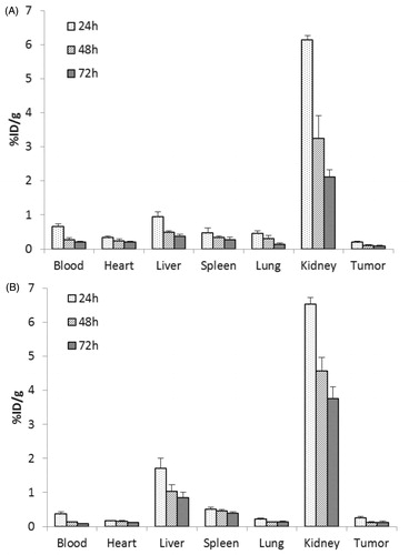
Table 3. The tumor-to-blood ratios of the 131I-labeled copolymers (n = 4).
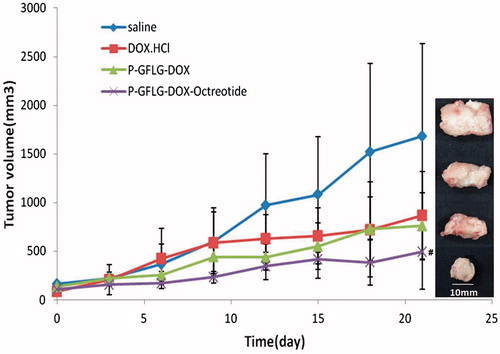
Figure 7. Antitumor activity of DOX·HCl, P-DOX and P-DOX-Oct on Kunming mice bearing H22 xenografts. The controls were administrated with saline. Tumor volume were recorded (n = 5, #p < 0.05, versus DOX·HCl) and tumors were harvested at the end of experiments (21 d).