1,233
Views
11
CrossRef citations to date
0
Altmetric
Research Article

Novel patient-specific navigational template for total knee arthroplasty

, , Ph.D., &
Pages 288-297 | Received 28 Dec 2010, Accepted 07 Jul 2011, Published online: 17 Oct 2011

Figures & data

Figure 1. 3D model of the knee. (a) Femoral view. (b) Tibial view.

Figure 1. 3D model of the knee. (a) Femoral view. (b) Tibial view.

Figure 2. Determining the optimal mechanical alignment of the femur (a), tibia (b), and medial tibial plateau line (c) using the Geomagic Studio10.0 software.
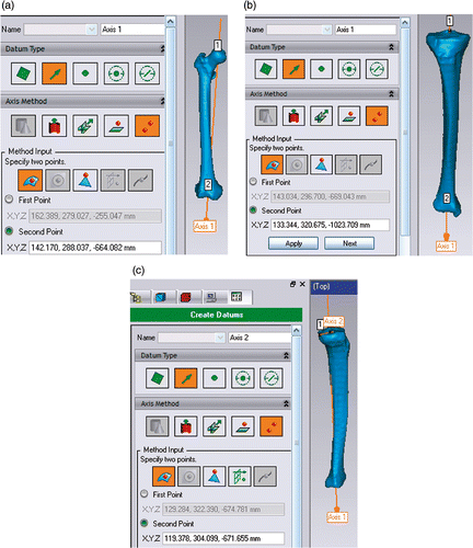
Figure 2. Determining the optimal mechanical alignment of the femur (a), tibia (b), and medial tibial plateau line (c) using the Geomagic Studio10.0 software.

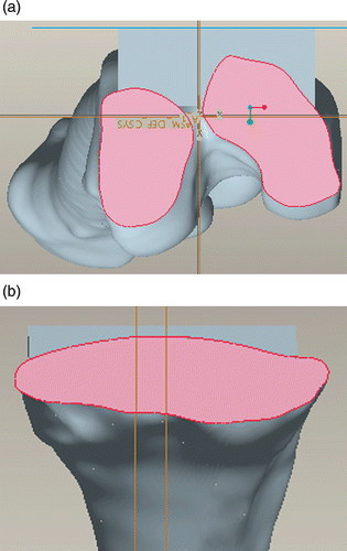
Figure 3. Design of the navigational templates. (a) Virtual femoral template. (b) Virtual tibial template.
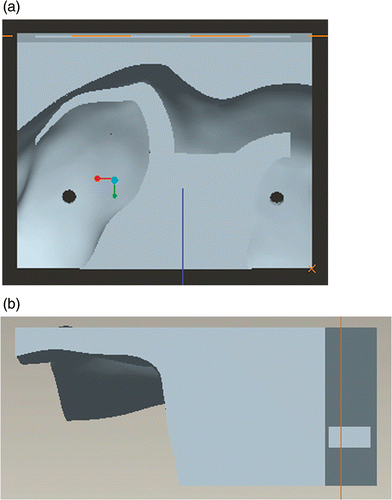
Figure 3. Design of the navigational templates. (a) Virtual femoral template. (b) Virtual tibial template.

Figure 4. Surface shape after virtually cutting the distal femur and proximal tibia.

Figure 4. Surface shape after virtually cutting the distal femur and proximal tibia.

Figure 5. The accuracy of the navigational template was examined by visual inspection. (a) RP model of the femoral condyle and tibial plateau. (b) and (c) The navigational template fits the RP model of the knee perfectly. (d) and (e) The gap for the osteotomy in the navigational template. (f) The rotational alignment for the femur defined by the two drill guides. (g) RP model of the distal femur and proximal tibia surfaces after osteotomy in the actual physical setting.
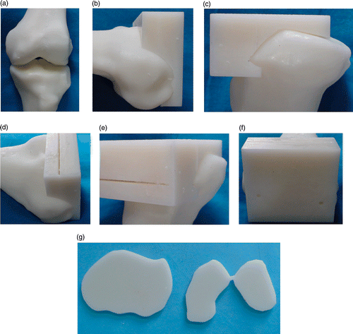
Figure 5. The accuracy of the navigational template was examined by visual inspection. (a) RP model of the femoral condyle and tibial plateau. (b) and (c) The navigational template fits the RP model of the knee perfectly. (d) and (e) The gap for the osteotomy in the navigational template. (f) The rotational alignment for the femur defined by the two drill guides. (g) RP model of the distal femur and proximal tibia surfaces after osteotomy in the actual physical setting.

Figure 6. Cadaver undergoing TKA using the patient-specific navigational template technique. (a) The navigational template fits the femoral condyle perfectly. (b) and (c) The actual surface of the distal femur after osteotomy matches the surface shape from the virtual setting using the navigational template. (d) The navigational template fits the tibial plateau perfectly. (e) and (f) The actual surface of the proximal tibia after osteotomy matches the surface shape from the virtual setting using the navigational template.
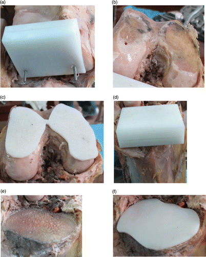
Figure 6. Cadaver undergoing TKA using the patient-specific navigational template technique. (a) The navigational template fits the femoral condyle perfectly. (b) and (c) The actual surface of the distal femur after osteotomy matches the surface shape from the virtual setting using the navigational template. (d) The navigational template fits the tibial plateau perfectly. (e) and (f) The actual surface of the proximal tibia after osteotomy matches the surface shape from the virtual setting using the navigational template.

Figure 7. Imaging examination showing good positioning of the prosthesis. (a) The resection of the posterior surface is parallel to the transepicondylar axis. (b) and (c) The distal resection of the femur and the proximal resection of the tibia are almost perpendicular to the mechanical axis of the leg in the coronal plane. (d) and (e) X-rays of the knee in anteroposterior and lateral views following TKA showed normal alignment of the knee with an intact prosthesis.
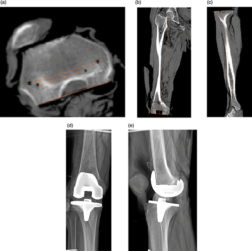
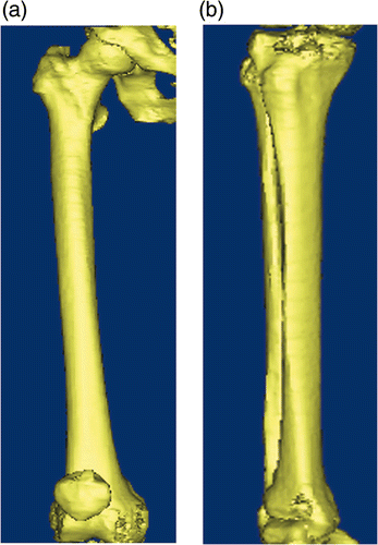
Figure 7. Imaging examination showing good positioning of the prosthesis. (a) The resection of the posterior surface is parallel to the transepicondylar axis. (b) and (c) The distal resection of the femur and the proximal resection of the tibia are almost perpendicular to the mechanical axis of the leg in the coronal plane. (d) and (e) X-rays of the knee in anteroposterior and lateral views following TKA showed normal alignment of the knee with an intact prosthesis.
Reprints and Corporate Permissions

Please note: Selecting permissions does not provide access to the full text of the article, please see our help page How do I view content?

To request a reprint or corporate permissions for this article, please click on the relevant link below:

Academic Permissions

Please note: Selecting permissions does not provide access to the full text of the article, please see our help page How do I view content?

Obtain permissions instantly via Rightslink by clicking on the button below:

If you are unable to obtain permissions via Rightslink, please complete and submit this Permissions form. For more information, please visit our Permissions help page.