Figures & data
Figure 1. Example of a SPME-GC chromatogram of human pericardial fat tissue that had been exposed to fixed temperatures or electrocautery, as indicated. Each peak indicates a volatile compound derived from FA decomposition and the amplitude its relative amount. The horizontal axis represents the retention time, during which the sample is gradually heated to allow evaporation of specific compounds. Compounds that evaporate at lower temperatures are seen to the left in each panel. Less volatile compounds require a higher temperature which is reached later during the run and are seen to the right along the retention-time axis. The peak at 12-min retention time refers to the internal standard used for output-signal calibration. Please, see Materials and Methods for further details.
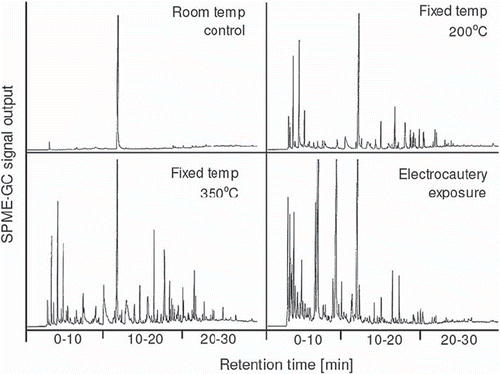
Figure 2. Peak counts on the SPME-GC chromatogram for pig fat exposed to different temperatures. The experiments are similar to the example shown for human fat in . For numeric data extraction the chromatograms were subdivided into three retention-time windows; 0–10, 10–20, and 20–30 min. The number of identifiable peaks was counted within each window. The peak referring to the internal standard at 12-min retention time was disregarded. A high count indicates a large number of compounds. Mean ± sem values, n=4. Please, see the legend to for explanatory details.
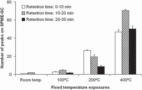
Table I. FA composition of human pericardial fat and the response to electrocautery.
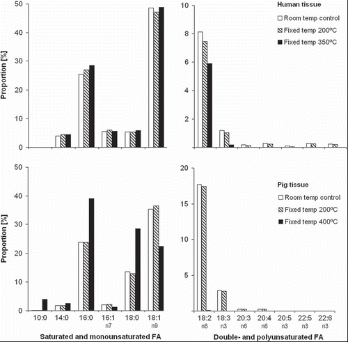
Figure 3. Fatty-acid (FA) proportions in pericardial tissue from humans and pigs in response to different temperature exposures, and with subdivision into FA saturation levels, as indicated. The data describe the overall FA composition of the sample. Fat samples were pooled for analysis, with n=3 and n=4 for human and pig tissue, respectively.