Figures & data
Figure 1. Representative electron microscopy of right atrium before (A) and during (B) coronary artery bypass grafting (CABG). Note injured and swollen mitochondria indicating ischemia and edema shown with arrows in B.
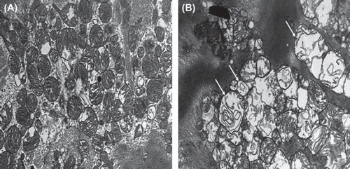
Figure 2. Syndecan-1 fold change concentrations adjusted to base line before coronary artery bypass grafting (Citation1), immediately after aortic declamping (Citation2) and after operation (Citation3) in controls (A, open circles) and in patients with Diazoxide (B, black triangles). Median is shown in each group with a horizontal line. An interpolation line has been added between median markers. Note significant increase (p < 0.004) and decrease (p < 0.04) from base line to time points 2 and 3, respectively, in patients with Diazoxide (B).
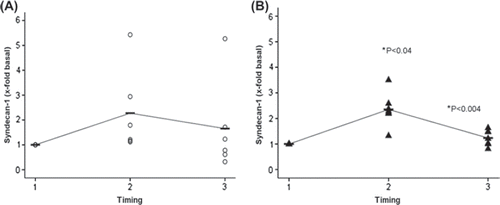
Figure 3. Hyaluronan fold change concentrations adjusted to base line before coronary artery bypass grafting (Citation1), immediately after aortic declamping (Citation2) and after operation (Citation3) in controls (A, open circles) and in patients with Diazoxide (B, black triangles). Median is shown in each group with a horizontal line. An interpolation line has been added between median markers. In both controls and patients with Diazoxide, an increase of Hyaluronan was noted at time point 2. Note significant decrease (p < 0.04) from time point 2 to 3 in patients with Diazoxide (B) but not controls (A).
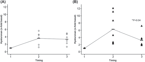
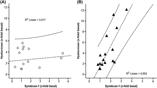
Figure 4. Correlation of Hyaluronan and syndecan-1 fold change values adjusted to base line for controls (A) and patients with Diazoxide (B). Note controlled correlation in B for patients with Diazoxide (R2 linear = 0.654, p < 0.005), but not for controls (R2 linear = 0.017, ns).