Figures & data
Figure 1. Structures of flavonoids.
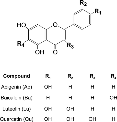
Figure 2. Effects of flavonoids on the mRNA expression of MMP-9 (A) and MMP-13 (B) in primary cultures of mouse osteoblasts. Cells were treated with 25 μM of each flavonoid, apigenin (Ap), bacalein (Ba), luteolin (Lu) and quercetin (Qu), for 1 h before IL-1β exposure. After further 24-h incubation, MMP-9 and -13 mRNA expressions were measured. *p < 0.05; **p < 0.01; ***p < 0.001 compared with IL-1β-treated control.
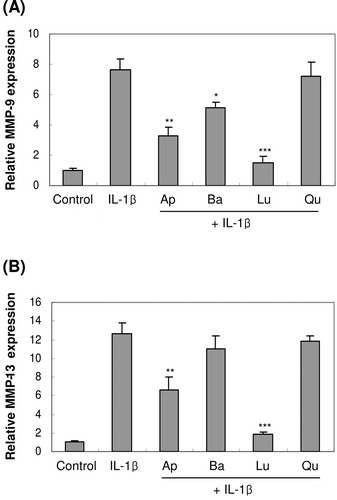
Figure 3. Concentration-dependent effect of luteolin on the expression of MMP-9 and -13 in primary cultures of osteoblasts. Cells were treated with luteolin at concentrations from 1.0 to 25 μM for 1 h before IL-1β exposure. After further 24-h incubation, MMP-9 and -13 mRNA expressions were measured. *p < 0.05; **p < 0.01; ***p < 0.001 compared with IL-1β-treated control.
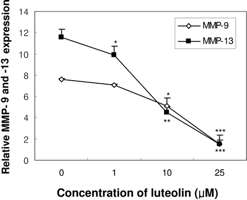
Figure 4. Effect of constitutively active form of the Ras protein (RasV12) and dominant-negative form of the Ras protein (RasN17) on ERK activation and the expression of MMP-9 and -13 mRNA in MC3T3-E1 cells. One day after the transfection with RasV12 or RasN17, cells were treated with IL-1β for 30 min for ERK phosphorylation (A) or treated for 24 h for the mRNA expression of MMP-9 (B) and MMP-13 (C). (CMV: empty vector, V: RasV12, N: RasN17). ##p < 0.01; ###p < 0.001 compared with control. **p < 0.01; ***p < 0.001 compared with IL-1β-treated control.
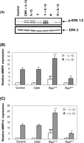
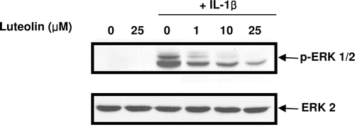
Figure 5. Effect of luteolin on IL-1β-induced ERK phosphorylation in primary cultures of mouse osteoblasts. Cells were pre-treated with luteolin at concentrations of 1, 10, 25 μM for 1 h before IL-1β exposure. After further 30-min incubation, ERK phosphorylation was measured using phosphor-specific ERK antibody.