Figures & data
Figure 1. The reactions catalysed by trypanothione reductase and glutathione reductase.
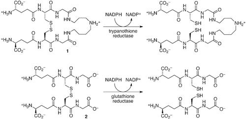
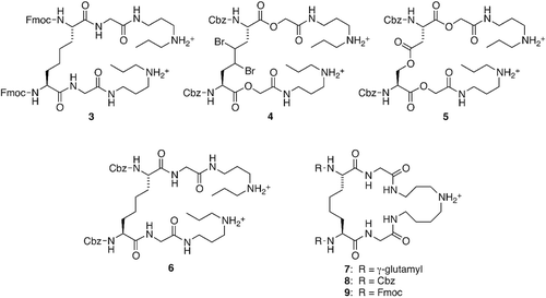
Figure 2. Inhibitors of trypanothione reductase.
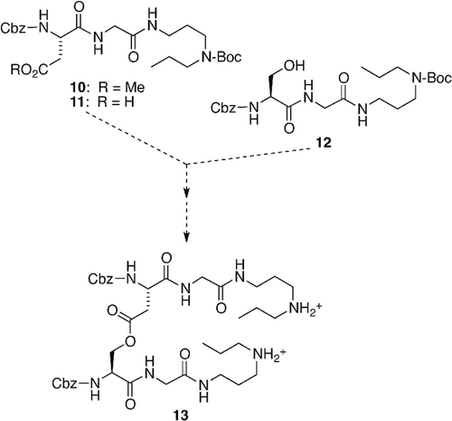
Scheme 1. Unsuccessful route to ester 13.
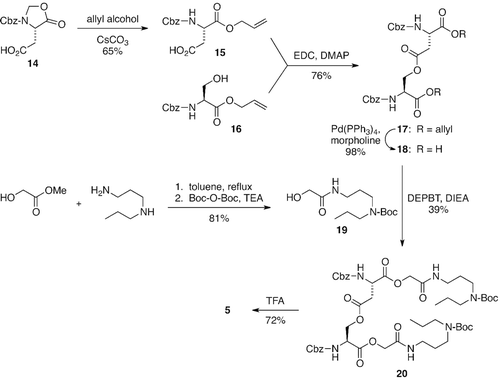
Scheme 2. The synthesis of inhibitor 5.
Scheme 3. The synthesis of inhibitors 3 and 4.
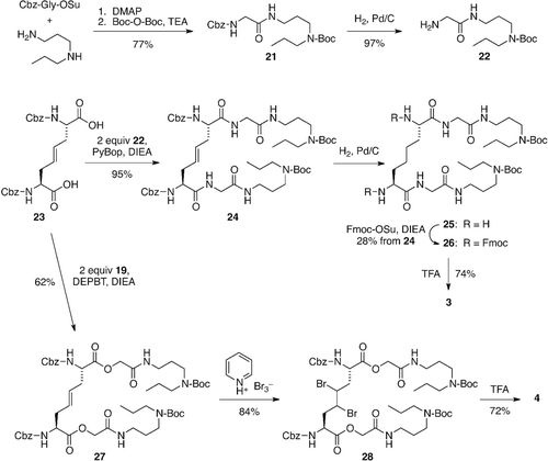
Table 1. Inhibition of TR by compounds 3–5 and by previously reported inhibitors 6–9a
Figure 3. TR assay substrate 29.
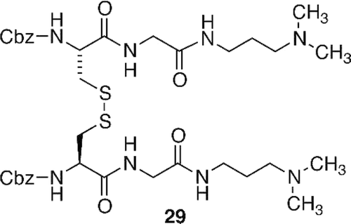
Figure 4. Competitive inhibition of TR y 3–5. (a) Lineweaver–Burk (L–B) plot for TR inhibition in the absence (•) and presence of 3 at 2.2 µM (□), 5.1 µM (▴), 10.2 µM (◊), 15.3 µM (▪), and 19.7 µM (○). (b) L–B plot for TR inhibition in the absence (•) and presence of 4 at 18.7 µM (□), 62.5 µM (▴), and 109.3 µM. (c) L–B plot for TR inhibition in the absence (•) and presence of 5 at 15.3 µM (□), 61.1 µM (▴), 107.0 µM (◊), and 152.9 µM (▪). Each point represents the average of two to four experimental determinations; the lines are based on theoretical values derived from the nonlinear least-squares fit of the data sets using the COMP program Citation31.
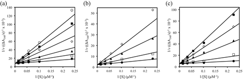
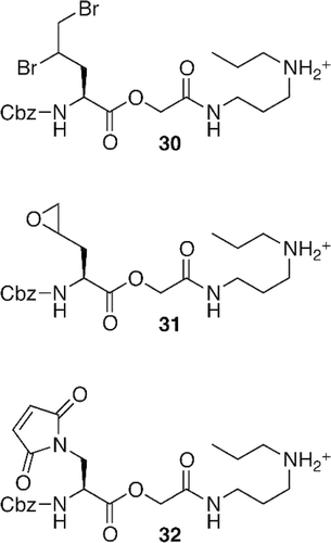
Figure 5. Potential electrophilic irreversible TR inhibitors.