Figures & data
Figure 1. Experimental design. (A) Timing of events. (B) Procedures performed on each group. Animals in all groups received middle cerebral artery occlusion (MCAO) and common carotid artery occlusion (CCAO) surgery under 1.5% isoflurane anesthesia. Immediately following focal cerebral ischemia, and depending on the assigned group, animals received 90 min of anaesthetic treatment or no treatment, and three intraperitoneal (IP) injections of either caspase-3 inhibitor (C3I) or dimethyl sulfoxide (DMSO) at day 0, 1, and 7 post-ischemia. Neurological evaluation was assessed in all groups at day 0, 4, and 14 post-ischemia. Animals in all groups were euthanized 14 days after focal cerebral ischemia, and brains were collected for analysis.
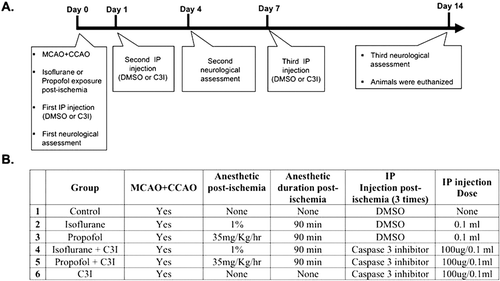
Table 1. Physiological Variables before and after Common Carotid Occlusion and Middle Cerebral Occlusion.
Figure 2. Neurological assessment using the eight-point behavioural rating scale. Behaviour measurements on post-MCAO day 4, showed a significantly better neurological outcome in both groups receiving a combination of an anaesthetic and a caspase 3 inhibitor (C3I). This trend is voided by post-day 14, when all animals approached baseline scores. Data are presented as mean ± SEM. *p < 0.05 versus control group at day 4.
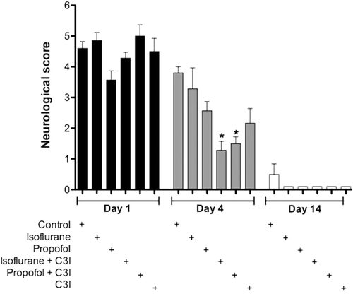
Figure 3. Cerebral infarction volume in each group measured 14 days following permanent focal ischemia. The graph shows the quantification of the average volume of the infarct 14 days after permanent focal cerebral ischemia as determined by stereological analysis of hematoxylin and eosin histochemistry. Data are presented as mean ± SEM. *p < 0.05 versus control group. C3I: Caspase 3 inhibitor.
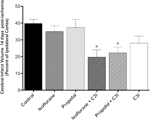
Figure 4. Estimated total number of TUNEL positive cells in the boundary area of cortical infarction 14 days post-ischemia. The number of TUNEL positive cells was significantly decreased in the isoflurane plus C3I, propofol plus C3I, and C3I alone as compared to the control group. Data are presented as mean ± SEM. *p < 0.05 versus control group. C3I: Caspase-3 inhibitor.
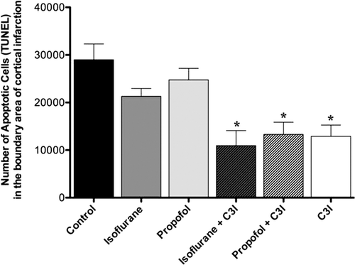
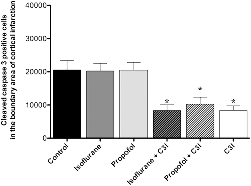
Figure 5. Estimated total number of cleaved caspase-3 positive cells in the boundary area of cortical infarction. The number of cleaved caspase-3 positive cells was significantly decreased in the animals that received isoflurane+ C3I, propofol + C3I and C3I alone as compared to the control group. Data are presented as mean ± SEM. *p < 0.05 versus control group. C3I: Caspase-3 inhibitor.