Figures & data
Figure 1. Sodium dodecyl sulphate-polyacrylamide gel electrophoresis (SDS-PAGE) analysis of purified LPO. Column 1: standard proteins: MBP (maltose-binding protein β-galactosidase, 175 kDa), MBP (maltose binding protein)-paramyosin (fusion of MBP and paramyosin, 80 kDa), maltose-binding protein and chitin binding domain (62 kDa), CBD-Mxe intein-2CBD (fusion of the chitin binding domain and the Mxe intein followed by two chitin-binding domains, 46 kDa). Column 2: purified LPO from bovine milk (80 kDa) (LPO: lactoperoxidase).
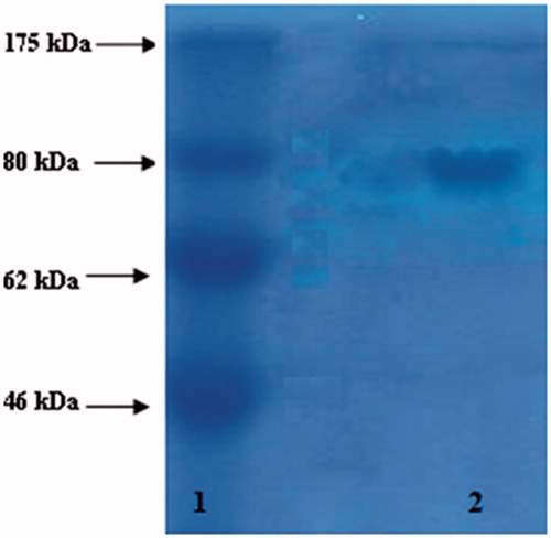
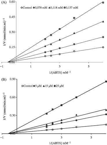
Figure 2. Lineweaver–Burk graph in five different substrate (ABTS) concentrations and in three different (A) ferulic acid and (B) quercetin concentrations for the determination of Ki.