Figures & data
Figure 1. Boxplot (median, interquartiles and range) of PCF in healthy controls and COPD patients according to GOLD grades.
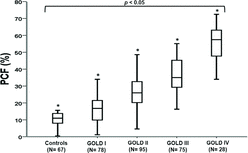
Figure 2. Significant correlations between% predicted total lung capacity (TLC) (panel A) and alveolar volume (VA) (panel B) with PCF in COPD patients grades 1 to 4 (N = 276). Panel C shows the non-linear relationship (asymptotic regression) between%TLC-%VA differences and PCF.
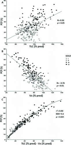
Figure 3. Non-linear correlations of PCF with FEV1, (panel A) and specific airway resistance (panel B) and linear correlations of PCF with residual volume (panel C) and lung diffusing capacity for carbon monoxide (panel D) in COPD patients grades 1 to 4 (N = 276). Lines represent the cutoffs for PCF tertiles.
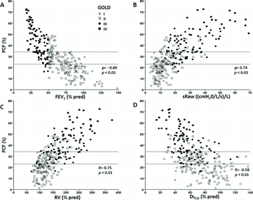
Figure 4. Absolute frequency of the main symptom (leg effort or shortness of breath (SOB) reported at the end of progressive exercise in COPD patients separated by PCF tertiles.
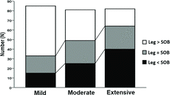
Table 1. Selected resting and peak exercise variables in COPD patients separated by PCF tertiles
Table 2. Selected resting and peak exercise variables in controls and COPD patients separated by PCF tertiles within GOLD grades
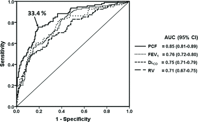
Figure 5. Receiver operating characteristics curve of selected physiological variables to predict a severely reduced peak exercise capacity in COPD patients GOLD grades 1 to 4 (N = 276). The best PCF cutoff is also highlighted. Definition of abbreviations: AUC: area under the curve; CI: confidence interval; PCF: poorly communicant fraction; FEV: forced expiratory volume in 1 second; DLCO: lung diffusing capacity for carbon monoxide; RV: residual volume.