828
Views
34
CrossRef citations to date
0
Altmetric
Research Article

Carbon monoxide form of PEGylated hemoglobin protects myocardium against ischemia/reperfusion injury in diabetic and normal mice

, , , , , , & show all
Pages 428-436 | Received 19 Dec 2012, Accepted 20 Dec 2012, Published online: 23 Jan 2013

Figures & data

Figure 1. Scheme depicting studies conducted in SANGUINATE-treated and SANGUINATE-untreated mice. (a) SANGUINATE was administered either prior to 30 min of LAD ligation and during 48 h of reperfusion or only during 48 h of reperfusion. (b) In the second set of experiments, mice were administered SANGUINATE only during 28 days of reperfusion after LAD ligation. Since PBS was used as a vehicle, in both (a) and (b), mice treated with PBS were used as the control group. (n = 12 in each group).

Figure 1. Scheme depicting studies conducted in SANGUINATE™-treated and SANGUINATE™-untreated mice. (a) SANGUINATE™ was administered either prior to 30 min of LAD ligation and during 48 h of reperfusion or only during 48 h of reperfusion. (b) In the second set of experiments, mice were administered SANGUINATE™ only during 28 days of reperfusion after LAD ligation. Since PBS was used as a vehicle, in both (a) and (b), mice treated with PBS were used as the control group. (n = 12 in each group).

Figure 2. Impact of myocardial infarction (MI) in SANGUINATE-treated diabetic and normal mice prior to LAD occlusion and during 48 h of reperfusion. Hearts retrieved after LAD ligation followed by 48 h of reperfusion and subjected to TTC staining (a) for the analysis of total area at risk (b) and infarcted area (c) in the following groups of mice: normal PBS-treated (NDM-PBS), SANGUINATE-treated NDM (NDM-Sang), diabetic PBS (DM-PBS), and SANGUINATE-treated DM (DM-Sang). (d) Troponin-C release in plasma, marker of ischemic injury, was also determined in all the groups of mice during the 48 h of reperfusion. NDM-Sang and DM-Sang mice received i.p. injections of SANGUINATE prior to LAD ligation and during 48 h of reperfusion. NDM-PBS and DM-PBS mice received i.p. injections of an equivalent volume of PBS vehicle prior to LAD ligation and during 48 h of reperfusion. (n = 12 in each group).

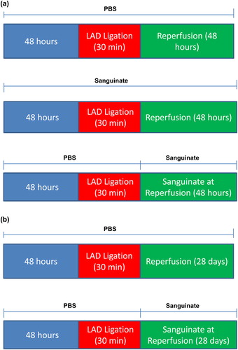
Figure 2. Impact of myocardial infarction (MI) in SANGUINATE™-treated diabetic and normal mice prior to LAD occlusion and during 48 h of reperfusion. Hearts retrieved after LAD ligation followed by 48 h of reperfusion and subjected to TTC staining (a) for the analysis of total area at risk (b) and infarcted area (c) in the following groups of mice: normal PBS-treated (NDM-PBS), SANGUINATE™-treated NDM (NDM-Sang), diabetic PBS (DM-PBS), and SANGUINATE™-treated DM (DM-Sang). (d) Troponin-C release in plasma, marker of ischemic injury, was also determined in all the groups of mice during the 48 h of reperfusion. NDM-Sang and DM-Sang mice received i.p. injections of SANGUINATE™ prior to LAD ligation and during 48 h of reperfusion. NDM-PBS and DM-PBS mice received i.p. injections of an equivalent volume of PBS vehicle prior to LAD ligation and during 48 h of reperfusion. (n = 12 in each group).

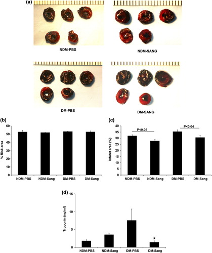
Table I. Cardiac Functional determined using echocardiography.

Figure 3. Impact of MI in SANGUINATE-treated diabetic and normal mice only during 48 h of reperfusion. Hearts retrieved after LAD ligation followed by 48 h of reperfusion and subjected to TTC staining (a) for the analysis of total area at risk (b) and infarcted area (c) in the myocardium were determined along with troponin-C (d) release in plasma during reperfusion. NDM-Sang and DM-Sang mice received i.p. injections of SANGUINATE only during 48 h of reperfusion. NDM-PBS and DM-PBS mice received i.p. injections of an equivalent volume of PBS vehicle during 48 h of reperfusion. (n = 12 in each group).

Figure 3. Impact of MI in SANGUINATE™-treated diabetic and normal mice only during 48 h of reperfusion. Hearts retrieved after LAD ligation followed by 48 h of reperfusion and subjected to TTC staining (a) for the analysis of total area at risk (b) and infarcted area (c) in the myocardium were determined along with troponin-C (d) release in plasma during reperfusion. NDM-Sang and DM-Sang mice received i.p. injections of SANGUINATE™ only during 48 h of reperfusion. NDM-PBS and DM-PBS mice received i.p. injections of an equivalent volume of PBS vehicle during 48 h of reperfusion. (n = 12 in each group).

Figure 4. Impact of MI in SANGUINATE-treated diabetic and normal mice only during 28 days of reperfusion. Hearts retrieved after LAD ligation followed by 28 days of reperfusion and subjected to TTC staining (a) for the analysis of total area at risk (b) and infarcted area (c) in the myocardium were determined. NDM-Sang and DM-Sang mice received i.p. injections of SANGUINATE only during 28 days of reperfusion. NDM-PBS and DM-PBS mice received i.p. injections of an equivalent volume of PBS vehicle during 48 h of reperfusion (n = 12 in each group).

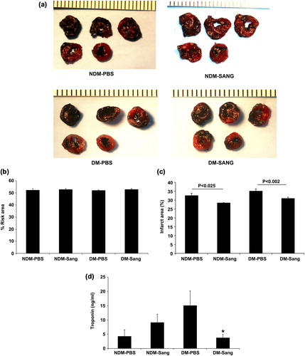
Figure 4. Impact of MI in SANGUINATE™-treated diabetic and normal mice only during 28 days of reperfusion. Hearts retrieved after LAD ligation followed by 28 days of reperfusion and subjected to TTC staining (a) for the analysis of total area at risk (b) and infarcted area (c) in the myocardium were determined. NDM-Sang and DM-Sang mice received i.p. injections of SANGUINATE™ only during 28 days of reperfusion. NDM-PBS and DM-PBS mice received i.p. injections of an equivalent volume of PBS vehicle during 48 h of reperfusion (n = 12 in each group).

Figure 5. Impact of SANGUINATE on oxidative stress changes in diabetic and normal mice after LAD ligation followed by reperfusion. Oxidative stress marker, malondialdehyde (MDA), was measured in heart lysates from NDM-PBS, NDM-Sang, DM-PBS, and DM-Sang in Study I (a) and Study II. Hearts in Studies I and II were harvested at the end of recovery period of 48 h. In Study I, mice were treated with SANGUINATE throughout LAD ligation and during 48 h of reperfusion (a), whereas in Study II the mice were treated with SANGUINATE only during 48 h of recovery after initiating reperfusion (b). More details on the protocols for Study I and Study II are described in the methods section. The MDA levels were determined using commercially available kits from Oxis. Data represent means ± SD; n = 4–6 per group.

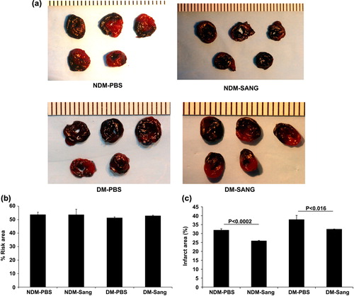
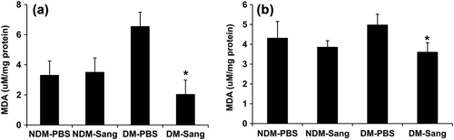
Figure 5. Impact of SANGUINATE™ on oxidative stress changes in diabetic and normal mice after LAD ligation followed by reperfusion. Oxidative stress marker, malondialdehyde (MDA), was measured in heart lysates from NDM-PBS, NDM-Sang, DM-PBS, and DM-Sang in Study I (a) and Study II. Hearts in Studies I and II were harvested at the end of recovery period of 48 h. In Study I, mice were treated with SANGUINATE™ throughout LAD ligation and during 48 h of reperfusion (a), whereas in Study II the mice were treated with SANGUINATE™ only during 48 h of recovery after initiating reperfusion (b). More details on the protocols for Study I and Study II are described in the methods section. The MDA levels were determined using commercially available kits from Oxis. Data represent means ± SD; n = 4–6 per group.

Reprints and Corporate Permissions

Please note: Selecting permissions does not provide access to the full text of the article, please see our help page How do I view content?

To request a reprint or corporate permissions for this article, please click on the relevant link below:

Academic Permissions

Please note: Selecting permissions does not provide access to the full text of the article, please see our help page How do I view content?

Obtain permissions instantly via Rightslink by clicking on the button below:

If you are unable to obtain permissions via Rightslink, please complete and submit this Permissions form. For more information, please visit our Permissions help page.