
Figures & data
Fig. 1 Characterization of microparticles (MPs) isolated from MCF-7/Dx cells. a) MPs isolated from MCF-7/Dx cells were analysed via flow cytometry and gated based on sized latex beads (0.3–1.1 µM); b) 34.4% of the gated MP population detected positive for P-gp expression using anti-P-gp mAb (BD Bioscience). All experiments were repeated at least 3 times with similar results. Data are representative of a typical experiment.
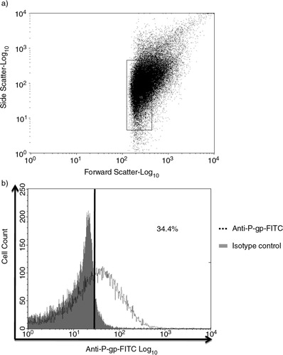
Fig. 2 Venn diagram depicting the comparison of proteins identified in microparticles (MPs). Proteins identified in MPs derived from drug-sensitive breast cancer cells (MCF-7) and drug-resistant breast cancer cells (MCF-7/Dx).
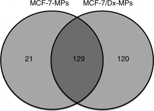
Fig. 3 Western blot analysis of selectively packaged proteins in MCF-7-Dx-MPs (microparticles). Twenty micrograms of total cell and MP lysates of drug-sensitive breast cancer cells (MCF-7) and drug-resistant breast cancer cells (MCF-7/Dx) were analysed by Western blot to detect (a) P-glycoprotein, 170 kDa; (b) ezrin, ~80 kDa; (c) moesin, 78–80 kDa; (d) CD44, 82 kDa; and (e) radixin, ~80 kDa. Lanes 1–4 are MCF-7 cells, MCF-7-MPs, MCF-7/Dx cells, and MCF-7/Dx-MPs, respectively. β-actin (42 kDa) was used as the internal control. Data are representative of a typical experiment.
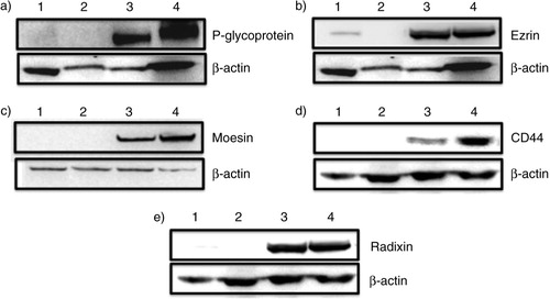
Table I Unique proteins identified in drug-resistant microparticles, which are correlated with cancer and/or multidrug resistance
Fig. 4 Protein network generated from String 9.05. Network of unique proteins associated with ABCB1/P-gp, isolated from microparticles derived from drug-resistant breast cancer cell (MCF-7/Dx), visualized on the String website. Proteins such as CD44, tropomyosin 3, glutathione S-transferase P1 and ecto-5′-nucleotidase are associated with drug-resistant proteins, whereas fibronectin 1, intercellular adhesion molecule 1, ezrin, moesin, integrin beta 3, vinculin, basigin, rho family and vimentin are associated with CD44.
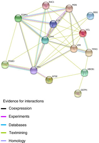
Fig. 5 Microparticles (MPs) play a deleterious role in cancer by transferring drug-resistant proteins. MP surface molecules (adhesion molecules) and FERM domain proteins are proposed to be associated with tissue selectivity in the transfer of P-gp in malignant breast cells.
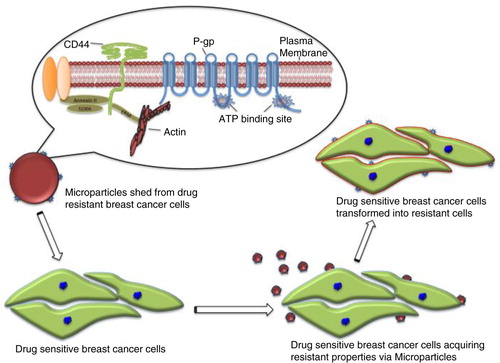
Table II Pathway analysis of proteins unique to resistant breast cancer–derived microparticles