Figures & data
Fig. 1 Physiological anticoagulant characteristics of blood vessels on endothelial cells. (a) Heparin-like proteoglycans located on endothelial cells’ surface enhance the inhibition capacity of anti-thrombin (AT). (b) Tissue factor pathway inhibitor (TFPI) inhibits tissue factor (TF) ability to initiate blood coagulation. TFPI actually binds to factor VIIa when this one interacts with TF. This inhibition is strengthened by the binding of factor Xa also neutralized by TFPI. (c) The protein C anticoagulant pathway takes place on endothelial cells surface. In this cascade, thrombin (T) procoagulant activity is inhibited by thrombomodulin (TM) enabling the activation of the protein C (PC) by thrombin via the endothelial cell protein C receptor (EPCR). The activated protein C (APC) detaches from the EPCR and interacts with the protein S (PS) to inactivate factor Va and VIIIa of the coagulation cascade.
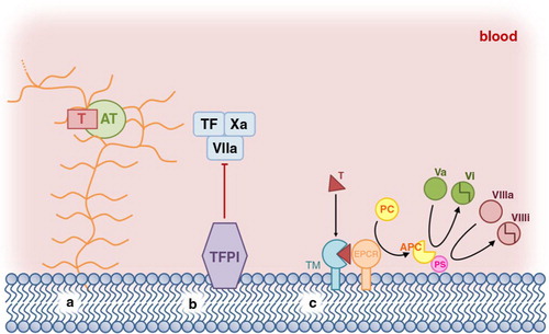
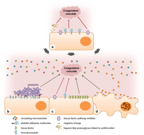
Fig. 2 Impact of extracellular vesicles on endothelial cells’ anticoagulant properties. (a) In a physiologic state, blood vessels’ endothelium exhibits anticoagulant properties. (b) Extracellular vesicles generated during tumourigenesis could increase platelet adhesion molecules’ expression by endothelial cells and thus, favour a procoagulant state of endothelium. (c) A decrease of tissue factor pathway inhibitor and thrombomodulin expression could be induced by extracellular vesicles. Extracellular vesicles could also trigger an increase in tissue factor on endothelial cells, contributing to a procoagulant phenotype. (d) Extracellular vesicles would induce endothelial cells’ apoptosis. Apoptotic endothelial cells exhibit negatively charged phospholipids and produce extracellular vesicles both contributing to a procoagulant phenotype of endothelial cells.