Figures & data
Figure 1. Different B cell functional response to inflammation.
Stimulation of any of the B cell functions depend on the nature of the pathogenic material, whereas memory B cells are long lasting immunological memory cells that bear specific receptors from previous infection.
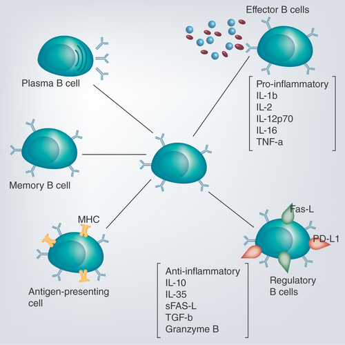
Figure 2. Biological pathways involved in development of regulatory B cells by various extracellular antigens have not yet been characterized and need further investigations.
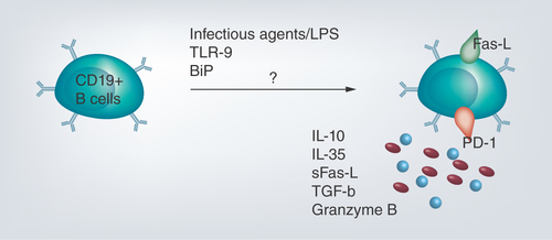
Figure 3. Circulating CD19+ B cells have the potential to develop to regulatory B cells with different regulatory markers depending on their stage of development.
The regulatory trait has been identified within B1 cells, marginal zone B cells and transitional-2 marginal zone B cells.
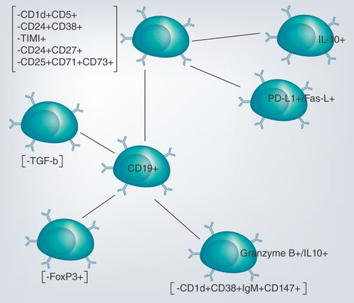
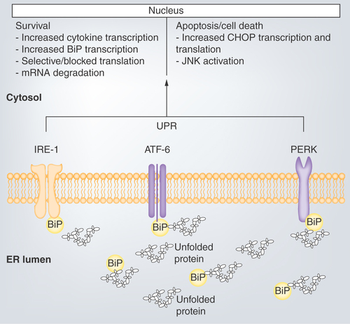
Figure 4. Unfolded protein response mediated by binding immunoglobulin protein.
Activation of the ER membrane transducers leads to a cascade of kinase phosphorylation that either favors apoptosis through upregulation of CHOP or JNK activation. Similarly, cell survival can be favored by blocking translation and degrading synthesized mRNA.
ER: Endoplasmic reticulum; UPR: Unfolded protein response.