Figures & data
Figure 1. Radiological findings of this case. (A) Abdominal computed tomography revealed multiple metastatic tumors in the liver and (B) thickening of the ascending colonic wall. (B, inset) Total colonoscopy demonstrated a large ulcerated and circumferential tumor at the same region.
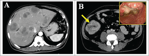
Figure 2. Immunohistochemical analysis of tumor cells. (A) The tumor cells were highly atypical small cells with hyperchromatic nuclei and scanty cytoplasm, and strongly positive for synaptophysin and Ki-67 (labeling index; 70%) (×400). (B) The tumor cells were positive for IGF-2, IGF-1R, and phosphorylated IGF-1R (×400).
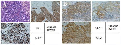
Table 1. Laboratory data on admission
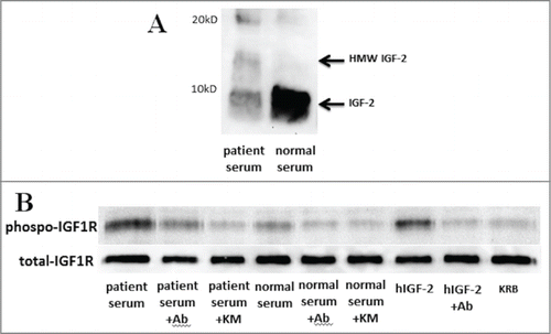
Figure 3. Western blot analysis and the kinase receptor activation assay of the patient's serum. (A) High molecular weight (HMW) and mature IGF-2 were detected in serum by western blot analysis using anti-IGF-2 antibody (ab9574). Each serum (40 μg of protein per lane) was separated by 15% SDS-PAGE. (B) IGF bioactivity in the patient's serum was greater than in a normal subject, and increased IGF bioactivity was inhibited to normal bioactivity level by the anti-IGF-2 specific antibody, ab9574, and increased IGF bioactivity was completely inhibited by the anti-IGF neutralizing antibody, KM1468. KRB; Krebs–Ringer bicarbonate buffer as a negative control, Ab; ab9574 (anti-IGF-2 specific antibody, final concentration 10 μg/ml), KM; KM1468 (anti-IGF neutralizing antibody, final concentration 10 μg/ml), hIGF-2; recombinant human IGF-2 (final concentration 10 ng/ml) as a positive control.