2,257
Views
18
CrossRef citations to date
0
Altmetric
Reports

Ciliary abnormalities in senescent human fibroblasts impair proliferative capacity

, , &
Pages 2773-2779 | Received 09 May 2014, Accepted 16 Jun 2014, Published online: 30 Oct 2014

Figures & data

Figure 1. Increased frequency and length of cilia in senescent human fibroblasts; (A) Doubling time over extended culture periods of BJ, MRC5 and NHDF cells; (B). Immunofluorescence microscopy of senescent BJ cells stained with antibodies to Cep135 (green) and γ-H2AX (red). DNA was visualised with DAPI (blue). Scale bar, 10 μm; (C) Quantitation of the frequency of centrosomal amplification in senescent BJ cells with γ-H2AX staining, scored as >4 centrin2 spots in a cell. Histograms show means ± s.d. of 3 separate experiments in which at least 200 cells were quantitated; (D) Quantitation of the ciliation frequency in the indicated cells, based on imaging of acetylated tubulin. ‘Unt’, untreated. Serum starvation (‘Ser.-stvd’) consisted of 24 h culture with 0.1% newborn calf serum. Histograms show means ± s.d. of 3 separate experiments in which at least 200 cells were quantitated; (E) Immunofluorescence microscopy of the indicated cells stained with antibodies to Cep135 (green) and acetylated tubulin (red). DNA was visualised with DAPI (blue). Scale bar, 10 μm; (F) Quantitation of cilium length in the indicated cell lines. At least 30 ciliated cells were scored for each. *, P < 0.05; **, P < 0.01; ***, P < 0.001 compared with controls of the same cell line by unpaired t-test.
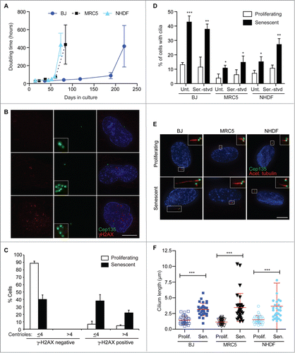
Figure 1. Increased frequency and length of cilia in senescent human fibroblasts; (A) Doubling time over extended culture periods of BJ, MRC5 and NHDF cells; (B). Immunofluorescence microscopy of senescent BJ cells stained with antibodies to Cep135 (green) and γ-H2AX (red). DNA was visualised with DAPI (blue). Scale bar, 10 μm; (C) Quantitation of the frequency of centrosomal amplification in senescent BJ cells with γ-H2AX staining, scored as >4 centrin2 spots in a cell. Histograms show means ± s.d. of 3 separate experiments in which at least 200 cells were quantitated; (D) Quantitation of the ciliation frequency in the indicated cells, based on imaging of acetylated tubulin. ‘Unt’, untreated. Serum starvation (‘Ser.-stvd’) consisted of 24 h culture with 0.1% newborn calf serum. Histograms show means ± s.d. of 3 separate experiments in which at least 200 cells were quantitated; (E) Immunofluorescence microscopy of the indicated cells stained with antibodies to Cep135 (green) and acetylated tubulin (red). DNA was visualised with DAPI (blue). Scale bar, 10 μm; (F) Quantitation of cilium length in the indicated cell lines. At least 30 ciliated cells were scored for each. *, P < 0.05; **, P < 0.01; ***, P < 0.001 compared with controls of the same cell line by unpaired t-test.

Figure 2. Cell-intrinsic control of extended cilium length in senescent cells; (A) Immunofluorescence microscopy of senescent BJ cells stained with antibodies to Arl13b (green) and γ-H2AX (red). DNA was visualised with DAPI (blue). Scale bar, 10 μm; (B) Quantitation of ciliation frequency in senescent BJ cells with γ-H2AX staining. Histograms show means ± s.d. of 3 separate experiments in which at least 200 cells were quantitated; (C) Immunofluorescence microscopy of senescent BJ cells stained with antibodies to centrin3 (red) and Arl13b (green) or Cep135 (red) and acetylated tubulin (green), as indicated. DNA was visualised with DAPI (blue). Scale bar, 10 μm; (D) Quantitation of cilium length in BJ cells, before or 24 h after the washout of 72 h 4 mM chloral hydrate treatment. This chloral hydrate treatment caused the removal of 90% of cilia from both proliferating and senescent cells. At least 30 ciliated cells were scored for each condition. **, P < 0.01; ***, P < 0.001 in comparison to the indicated controls by unpaired t-test; (E) Immunoblot analysis of CP110 expression in BJ cells. Size markers are indicated in kDa.
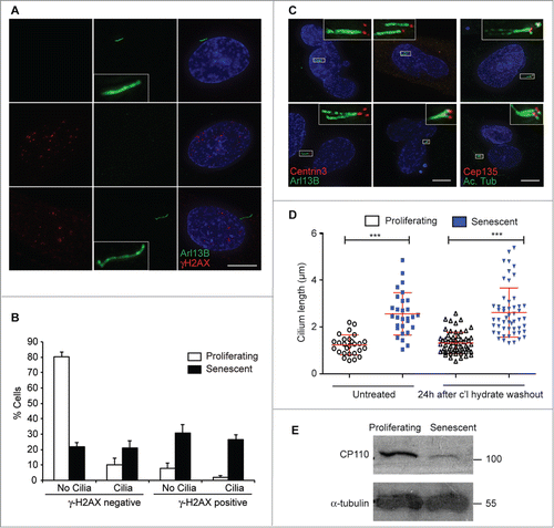
Figure 2. Cell-intrinsic control of extended cilium length in senescent cells; (A) Immunofluorescence microscopy of senescent BJ cells stained with antibodies to Arl13b (green) and γ-H2AX (red). DNA was visualised with DAPI (blue). Scale bar, 10 μm; (B) Quantitation of ciliation frequency in senescent BJ cells with γ-H2AX staining. Histograms show means ± s.d. of 3 separate experiments in which at least 200 cells were quantitated; (C) Immunofluorescence microscopy of senescent BJ cells stained with antibodies to centrin3 (red) and Arl13b (green) or Cep135 (red) and acetylated tubulin (green), as indicated. DNA was visualised with DAPI (blue). Scale bar, 10 μm; (D) Quantitation of cilium length in BJ cells, before or 24 h after the washout of 72 h 4 mM chloral hydrate treatment. This chloral hydrate treatment caused the removal of 90% of cilia from both proliferating and senescent cells. At least 30 ciliated cells were scored for each condition. **, P < 0.01; ***, P < 0.001 in comparison to the indicated controls by unpaired t-test; (E) Immunoblot analysis of CP110 expression in BJ cells. Size markers are indicated in kDa.

Figure 3. Hedgehog inhibition blocks proliferation and increases cilium length; (A) Immunofluorescence microscopy of BJ cells stained with antibodies to Ki67 (green). DNA was visualised with DAPI (blue). Scale bar, 10 μm; (B) Quantitation of the proliferative index of BJ cells after the indicated treatment, as determined by microscopy analysis of Ki67 signal. Histograms show means ± s.d. of 3 separate experiments in which at least 100 cells were quantitated; (C) Quantitation of cilium length in the indicated cells. At least 30 ciliated cells were scored for each condition, even when there were very few such cells. ***, P < 0.001 in comparison to untreated or indicated controls by unpaired t-test. ‘ns’, not significant; (D) Immunoblot analysis of the indicated protein expression in BJ cells. Size markers are indicated in kDa.
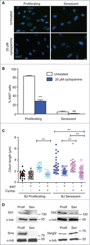
Figure 3. Hedgehog inhibition blocks proliferation and increases cilium length; (A) Immunofluorescence microscopy of BJ cells stained with antibodies to Ki67 (green). DNA was visualised with DAPI (blue). Scale bar, 10 μm; (B) Quantitation of the proliferative index of BJ cells after the indicated treatment, as determined by microscopy analysis of Ki67 signal. Histograms show means ± s.d. of 3 separate experiments in which at least 100 cells were quantitated; (C) Quantitation of cilium length in the indicated cells. At least 30 ciliated cells were scored for each condition, even when there were very few such cells. ***, P < 0.001 in comparison to untreated or indicated controls by unpaired t-test. ‘ns’, not significant; (D) Immunoblot analysis of the indicated protein expression in BJ cells. Size markers are indicated in kDa.

Figure 4. CP110 depletion causes increased ciliation and reduced cellular proliferation; (A) Quantitation of CP110 depletion in BJ cells by siRNA as determined by quantitative RT-PCR. Knockdowns are compared to the effect of GAPDH siRNA normalized to housekeeping gene expression. Data show the mean—s.d. of 3 separate experiments; (B) Quantitation of the ciliation frequency in BJ cells after the indicated treatments. Histograms show means + s.d. of 3 separate experiments in which at least 100 cells were quantitated; (C) Microscopy of CP110 siRNA-transfected cells stained with antibodies to Ki67 (red in merge). Fluorescent RNA (FAM; green) was cotransfected at a ratio of 1:5 with the siRNA as a transfection control. DNA was visualised with DAPI (blue). Scale bar, 10 μm; (D) Quantitation of the proliferative index of BJ cells after the indicated treatment, as determined by microscopy analysis of Ki67 signal. Histograms show means ± s.d. of 3 separate experiments in which at least 100 transfected cells were quantitated; *, P < 0.05; **, P < 0.01 in comparison with GAPDH siRNA controls by unpaired t-test.
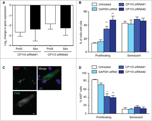
Figure 4. CP110 depletion causes increased ciliation and reduced cellular proliferation; (A) Quantitation of CP110 depletion in BJ cells by siRNA as determined by quantitative RT-PCR. Knockdowns are compared to the effect of GAPDH siRNA normalized to housekeeping gene expression. Data show the mean—s.d. of 3 separate experiments; (B) Quantitation of the ciliation frequency in BJ cells after the indicated treatments. Histograms show means + s.d. of 3 separate experiments in which at least 100 cells were quantitated; (C) Microscopy of CP110 siRNA-transfected cells stained with antibodies to Ki67 (red in merge). Fluorescent RNA (FAM; green) was cotransfected at a ratio of 1:5 with the siRNA as a transfection control. DNA was visualised with DAPI (blue). Scale bar, 10 μm; (D) Quantitation of the proliferative index of BJ cells after the indicated treatment, as determined by microscopy analysis of Ki67 signal. Histograms show means ± s.d. of 3 separate experiments in which at least 100 transfected cells were quantitated; *, P < 0.05; **, P < 0.01 in comparison with GAPDH siRNA controls by unpaired t-test.
Supplemental material

Supplemental_Files

Download Zip (3.9 MB)

Reprints and Corporate Permissions

Please note: Selecting permissions does not provide access to the full text of the article, please see our help page How do I view content?

To request a reprint or corporate permissions for this article, please click on the relevant link below:

Academic Permissions

Please note: Selecting permissions does not provide access to the full text of the article, please see our help page How do I view content?

Obtain permissions instantly via Rightslink by clicking on the button below:

If you are unable to obtain permissions via Rightslink, please complete and submit this Permissions form. For more information, please visit our Permissions help page.