985
Views
11
CrossRef citations to date
0
Altmetric
RESEARCH PAPERS

Candida albicansVPS4 contributes differentially to epithelial and mucosal pathogenesis

, , , , &
Pages 810-818 | Received 14 Apr 2014, Accepted 17 Aug 2014, Published online: 31 Oct 2014

Figures & data

Figure 1. The C. albicans vps4Δ mutant is more sensitive to cell wall stress and challenge with antifungals. C. albicans DAY185, vps4Δ, and VPS4 reintegrant strains were grown on agar plates containing cell-wall perturbing agents or antifungal drugs. Strains are indicated on the left. Cell densities decrease from left to right (1 × 10⁷, 2 × 10⁶, 4 × 10⁵, and 8 × 10⁴ cells per mL). Normal growth on YPD medium is shown on the top right. YPD medium containing cell wall perturbing agents including 0.02% SDS, 200 μg/mL Congo Red, and 50 μg/mL Calcofluor White, as well as medium containing the antifungal drugs caspofungin (0.1 μg/mL) and fluconazole (4 μg/mL), are shown.
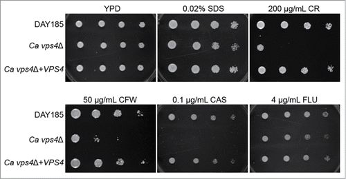
Figure 1. The C. albicans vps4Δ mutant is more sensitive to cell wall stress and challenge with antifungals. C. albicans DAY185, vps4Δ, and VPS4 reintegrant strains were grown on agar plates containing cell-wall perturbing agents or antifungal drugs. Strains are indicated on the left. Cell densities decrease from left to right (1 × 10⁷, 2 × 10⁶, 4 × 10⁵, and 8 × 10⁴ cells per mL). Normal growth on YPD medium is shown on the top right. YPD medium containing cell wall perturbing agents including 0.02% SDS, 200 μg/mL Congo Red, and 50 μg/mL Calcofluor White, as well as medium containing the antifungal drugs caspofungin (0.1 μg/mL) and fluconazole (4 μg/mL), are shown.

Figure 2. The C. albicans vps4Δ mutant is attenuated in macrophage killing. C. albicans cells were co-incubated with macrophage cells in unbuffered DMEM+5%FCS at an MOI of 2. (A) Counts of live macrophage cells from 12 separate fields after 24 h of co-incubation with C. albicans strains. Asterisk (*) denotes statistical significance, P < 0.05, compared to all other treatments. Experiment was performed in quadruplicate; the average of all 4 experiments is shown. (B) Live (green) and dead (red) macrophage cells were co-stained with calcein AM and ethidium bromide homodimer, respectively, and visualized by fluorescence microscopy. Representative images from the 24 h timepoint are shown.
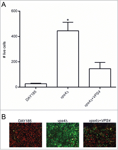
Figure 2. The C. albicans vps4Δ mutant is attenuated in macrophage killing. C. albicans cells were co-incubated with macrophage cells in unbuffered DMEM+5%FCS at an MOI of 2. (A) Counts of live macrophage cells from 12 separate fields after 24 h of co-incubation with C. albicans strains. Asterisk (*) denotes statistical significance, P < 0.05, compared to all other treatments. Experiment was performed in quadruplicate; the average of all 4 experiments is shown. (B) Live (green) and dead (red) macrophage cells were co-stained with calcein AM and ethidium bromide homodimer, respectively, and visualized by fluorescence microscopy. Representative images from the 24 h timepoint are shown.

Figure 3. The C. albicans vps4Δ mutant is attenuated in virulence in a Caenorhabditis elegans model of infection. C. albicans DAY185, vps4Δ, and the VPS4 reintegrant were tested in a C. elegans model of intestinal epithelial infection. C. elegans N2 nematodes were incubated with C. albicans cells for 4 hours, then monitored twice daily for survival. (A) Survival curve of C. elegans nematodes infected with C. albicans strains of interest. Survival of nematodes infected with C. albicans DAY185, vps4Δ, or VPS4 reintegrant cells, or fed nonpathogenic E. coli OP50 as a negative control, is shown as percent survival relative to day zero. (B) Light microscopy of nematodes killed by C. albicans DAY185, vps4Δ, or VPS4 reintegrant. After worms were scored as dead, worms were visualized via light microscopy. Extensive hyphae are seen piercing the cuticle of worms infected with C. albicans DAY185 and VPS4 reintegrant cells. Less extensive hyphae (indicated by black arrows) are visible protruding from the intestine of worms infected with the C. albicans vps4Δ null mutant.
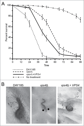
Figure 3. The C. albicans vps4Δ mutant is attenuated in virulence in a Caenorhabditis elegans model of infection. C. albicans DAY185, vps4Δ, and the VPS4 reintegrant were tested in a C. elegans model of intestinal epithelial infection. C. elegans N2 nematodes were incubated with C. albicans cells for 4 hours, then monitored twice daily for survival. (A) Survival curve of C. elegans nematodes infected with C. albicans strains of interest. Survival of nematodes infected with C. albicans DAY185, vps4Δ, or VPS4 reintegrant cells, or fed nonpathogenic E. coli OP50 as a negative control, is shown as percent survival relative to day zero. (B) Light microscopy of nematodes killed by C. albicans DAY185, vps4Δ, or VPS4 reintegrant. After worms were scored as dead, worms were visualized via light microscopy. Extensive hyphae are seen piercing the cuticle of worms infected with C. albicans DAY185 and VPS4 reintegrant cells. Less extensive hyphae (indicated by black arrows) are visible protruding from the intestine of worms infected with the C. albicans vps4Δ null mutant.

Figure 4. The C. albicans vps4Δ mutant causes less LDH release by oral epithelial cells in an oral epithelial model of infection. C. albicans DAY185, vps4Δ, and the VPS4 reintegrant were tested in an oral epithelial model of tissue invasion. C. albicans cells were co-incubated with oral epithelial cells for 24 h and then assayed for lactate dehydrogenase (LDH) release. LDH units were calculated by extrapolation from a standard curve and the number of LDH units released by the untreated positive control was subtracted from the total LDH units released by the infected oral epithelial cells.
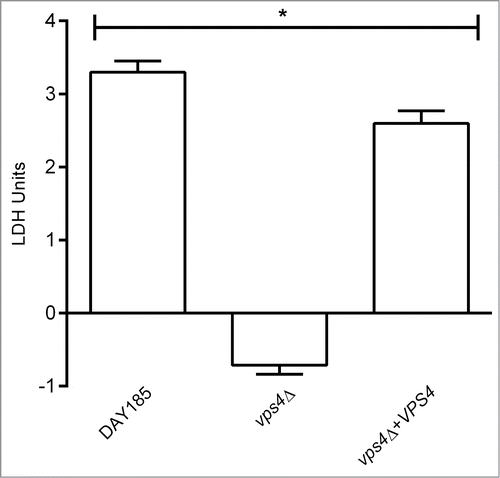
Figure 4. The C. albicans vps4Δ mutant causes less LDH release by oral epithelial cells in an oral epithelial model of infection. C. albicans DAY185, vps4Δ, and the VPS4 reintegrant were tested in an oral epithelial model of tissue invasion. C. albicans cells were co-incubated with oral epithelial cells for 24 h and then assayed for lactate dehydrogenase (LDH) release. LDH units were calculated by extrapolation from a standard curve and the number of LDH units released by the untreated positive control was subtracted from the total LDH units released by the infected oral epithelial cells.

Figure 5. The C. albicans vps4Δ mutant is less adherent to uro-epithelial cells in a uro-epithelial model of infection. C. albicans DAY185, vps4Δ, and the VPS4 reintegrant were tested in a monolayer uro-epithelial model. (A) Adherence of C. albicans cells to the uro-epithelial monolayer after 2 h and 24 h of co-incubation was assessed by CFU counts. Asterisk (*) denotes statistical significance, P < 0.05, compared to all other treatments. (B) C. albicans cells were co-incubated with uro-epithelial cells for 2 h and 24 h and then assayed for lactate dehydrogenase (LDH) release.% LDH release was calculated as a percentage as compared to a maximum LDH release control.
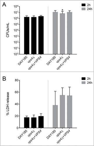
Figure 5. The C. albicans vps4Δ mutant is less adherent to uro-epithelial cells in a uro-epithelial model of infection. C. albicans DAY185, vps4Δ, and the VPS4 reintegrant were tested in a monolayer uro-epithelial model. (A) Adherence of C. albicans cells to the uro-epithelial monolayer after 2 h and 24 h of co-incubation was assessed by CFU counts. Asterisk (*) denotes statistical significance, P < 0.05, compared to all other treatments. (B) C. albicans cells were co-incubated with uro-epithelial cells for 2 h and 24 h and then assayed for lactate dehydrogenase (LDH) release.% LDH release was calculated as a percentage as compared to a maximum LDH release control.

Figure 6. The C. albicans vps4Δ mutant is virulent in a mouse model of vaginal candidiasis. C. albicans DAY185, vps4Δ, and the VPS4 reintegrant were tested for virulence in a mouse model of vaginal candidiasis. Mice were infected with 5 × 10⁵ C. albicans cells or with a PBS-only control. (A) Fungal burden after 3, 7 and 10 days of C. albicans infection. Fungal burden is indicated by CFUs per mL. (B) Vaginal histopathology comparing tissue invasion and damage caused by (a) wild-type SC5314, (b) wild-type DAY185, (c) vps4Δ and (d) the VPS4 reintegrant strain. Paraffin sections of vaginas from randomly selected mice were prepared and stained with hematoxylin.
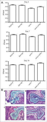
Figure 6. The C. albicans vps4Δ mutant is virulent in a mouse model of vaginal candidiasis. C. albicans DAY185, vps4Δ, and the VPS4 reintegrant were tested for virulence in a mouse model of vaginal candidiasis. Mice were infected with 5 × 10⁵ C. albicans cells or with a PBS-only control. (A) Fungal burden after 3, 7 and 10 days of C. albicans infection. Fungal burden is indicated by CFUs per mL. (B) Vaginal histopathology comparing tissue invasion and damage caused by (a) wild-type SC5314, (b) wild-type DAY185, (c) vps4Δ and (d) the VPS4 reintegrant strain. Paraffin sections of vaginas from randomly selected mice were prepared and stained with hematoxylin.

Reprints and Corporate Permissions

Please note: Selecting permissions does not provide access to the full text of the article, please see our help page How do I view content?

To request a reprint or corporate permissions for this article, please click on the relevant link below:

Academic Permissions

Please note: Selecting permissions does not provide access to the full text of the article, please see our help page How do I view content?

Obtain permissions instantly via Rightslink by clicking on the button below:

If you are unable to obtain permissions via Rightslink, please complete and submit this Permissions form. For more information, please visit our Permissions help page.