Figures & data
Figure 1. Schematic description of the protocol used to detect adenosine by using an aptamer-based gold nanoparticle enhanced SPR protocol. Reproduced with permission by the American Chemical Society.Citation41
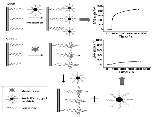
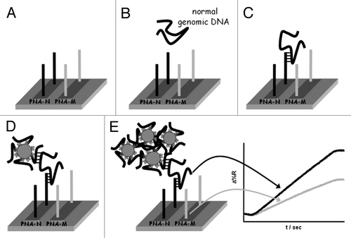
Figure 2. Pictorial description of the nanoparticle-enhanced SPRI strategy used to detect the normal, heterozygous and homozygous genomic DNAs. PNA-N and PNA-M specifically recognize the normal β-globin and the mutated β°39-globin genomic sequences respectively. Reproduced with permission by the American Chemical Society.Citation89