1,755
Views
44
CrossRef citations to date
0
Altmetric
Translational Brief Report

ATRA-induced upregulation of Beclin 1 prolongs the life span of differentiated acute promyelocytic leukemia cells

, , , , , , & show all
Pages 1108-1114 | Received 23 Sep 2010, Accepted 08 Jun 2011, Published online: 01 Oct 2011

Figures & data

Figure 1 ATRA activates autophagic flux in APL cells NB4 cells (1 × 10⁵ cells/ml) were treated with ATRA (1 µM) for the times indicated in the presence and absence of E64 (10 µg/ml) + methyl ester pepstatin (1 µg/ml). (A) Cells were examined by electron microscopy during the time course of ATRA-induced granulocytic differentiation. At time 0, the cells displayed rounded nuclei filled with euchromatin, and a ribosome-rich cytoplasm with well-defined mitochondria. After 4 and 6 d of ATRA treatment, the nuclei were poly-lobed and had a higher heterochromatin content. Most of the cell profiles also contained several prominent cytoplasmic vacuoles containing large amounts of membranous structures at different stages of degradation (arrows). (B) Autophagy and autophagic flux were also evaluated by immunoblot analysis of LC3-II levels in cell extracts, using β actin as a loading control. (C) Beclin 1 and Bcl-2 protein levels and mTOR activity were determined following immunoblot analysis using antibodies directed against Beclin 1, Bcl-2, phospho-p70S6 kinase, p70s6 kinase and β actin. (D) Immunoblot analysis of LC3-II and Beclin 1 proteins in HL60 cells incubated with ATRA for 3 or 4 d.
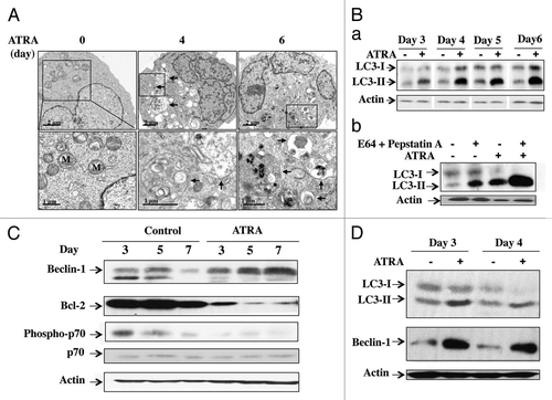
Figure 1 ATRA activates autophagic flux in APL cells NB4 cells (1 × 10⁵ cells/ml) were treated with ATRA (1 µM) for the times indicated in the presence and absence of E64 (10 µg/ml) + methyl ester pepstatin (1 µg/ml). (A) Cells were examined by electron microscopy during the time course of ATRA-induced granulocytic differentiation. At time 0, the cells displayed rounded nuclei filled with euchromatin, and a ribosome-rich cytoplasm with well-defined mitochondria. After 4 and 6 d of ATRA treatment, the nuclei were poly-lobed and had a higher heterochromatin content. Most of the cell profiles also contained several prominent cytoplasmic vacuoles containing large amounts of membranous structures at different stages of degradation (arrows). (B) Autophagy and autophagic flux were also evaluated by immunoblot analysis of LC3-II levels in cell extracts, using β actin as a loading control. (C) Beclin 1 and Bcl-2 protein levels and mTOR activity were determined following immunoblot analysis using antibodies directed against Beclin 1, Bcl-2, phospho-p70S6 kinase, p70s6 kinase and β actin. (D) Immunoblot analysis of LC3-II and Beclin 1 proteins in HL60 cells incubated with ATRA for 3 or 4 d.

Figure 2 Beclin 1 prolongs the life span of mature APL cells. NB4 cells were transfected with control or BECN1-specific siRNAs and then treated with ATRA (1 µM) for the times indicated. (A) Immunoblot analysis of Beclin 1 and LC3-II in NB4 cells incubated with either ATRA for 3 d or nutrient-free (NF) medium for 3 h. (B) The percentage of cells carrying apoptotic nuclei was determined by Hoechst staining at the times indicated. (C) The mitochondrial transmembrane potential was assessed using TMRM, as described in Materials and Methods, in NB4 cells treated with ATRA for 4 d; M represents the population of cells with low TMRM staining. (D) The cleavage of caspase-3 was examined by protein gel blotting using an antibody against caspase-3 in NB4 cells treated with ATRA for 3 d.
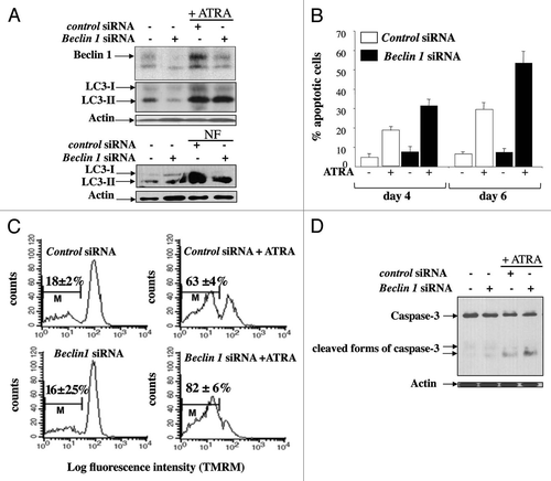
Figure 2 Beclin 1 prolongs the life span of mature APL cells. NB4 cells were transfected with control or BECN1-specific siRNAs and then treated with ATRA (1 µM) for the times indicated. (A) Immunoblot analysis of Beclin 1 and LC3-II in NB4 cells incubated with either ATRA for 3 d or nutrient-free (NF) medium for 3 h. (B) The percentage of cells carrying apoptotic nuclei was determined by Hoechst staining at the times indicated. (C) The mitochondrial transmembrane potential was assessed using TMRM, as described in Materials and Methods, in NB4 cells treated with ATRA for 4 d; M represents the population of cells with low TMRM staining. (D) The cleavage of caspase-3 was examined by protein gel blotting using an antibody against caspase-3 in NB4 cells treated with ATRA for 3 d.

Figure 3 Effect of silencing BECN1 expression on granulocyte differentiation in APL cells. (A–C) NB4 cells were transfected with control or BECN1-specific siRNAs, and then treated with ATRA (1 µM) for the times indicated. Granulocytic differentiation was assessed by: (A), measuring CD11c expression by FACS after 3 and 4 d of ATRA treatment; M is the percentage of cells expressing high levels of CD11c. (B) May-Grunwald Giemsa staining after 4 d of ATRA treatment; inset, the levels of Beclin 1 protein expression in ATRA-treated NB4 cells in the presence and absence of BECN1 siRNA are shown. (C) Quantification of anion superoxide production by using dihydroethidium (HE) dye in NB4 cells treated with ATRA for 2 or 3 d. Results are expressed as the ratio of the population of cells with high HE staining in ATRA-treated cells to the population of cells with high HE in control cells.
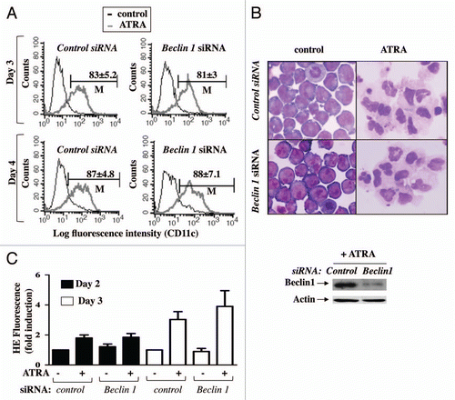
Figure 3 Effect of silencing BECN1 expression on granulocyte differentiation in APL cells. (A–C) NB4 cells were transfected with control or BECN1-specific siRNAs, and then treated with ATRA (1 µM) for the times indicated. Granulocytic differentiation was assessed by: (A), measuring CD11c expression by FACS after 3 and 4 d of ATRA treatment; M is the percentage of cells expressing high levels of CD11c. (B) May-Grunwald Giemsa staining after 4 d of ATRA treatment; inset, the levels of Beclin 1 protein expression in ATRA-treated NB4 cells in the presence and absence of BECN1 siRNA are shown. (C) Quantification of anion superoxide production by using dihydroethidium (HE) dye in NB4 cells treated with ATRA for 2 or 3 d. Results are expressed as the ratio of the population of cells with high HE staining in ATRA-treated cells to the population of cells with high HE in control cells.

Reprints and Corporate Permissions

Please note: Selecting permissions does not provide access to the full text of the article, please see our help page How do I view content?

To request a reprint or corporate permissions for this article, please click on the relevant link below:

Academic Permissions

Please note: Selecting permissions does not provide access to the full text of the article, please see our help page How do I view content?

Obtain permissions instantly via Rightslink by clicking on the button below:

If you are unable to obtain permissions via Rightslink, please complete and submit this Permissions form. For more information, please visit our Permissions help page.