Figures & data
Figure 1. Schematic of the fabrication of PLGA core + pNIPAM shell nanoparticles (A and B) and the subsequent chemistry (C) used to append the collagen type II binding peptide targeting ligand.
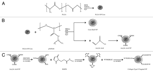
Table 1. Characterization of Various Nanoparticle Samples
Figure 2. Transmission electron microscope images of the various nanoparticles both pre- and post-lyophilization. Scale bars = 250 nm.
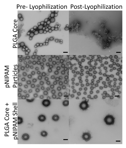
Figure 3. Temperature sweeps of the various nanoparticles. Data are represented as mean ± standard deviation (n = 3). C, PLGA core only; C+S #%, PLGA core + pNIPAM shell + mol% acrylic acid; L, Lyophilized.
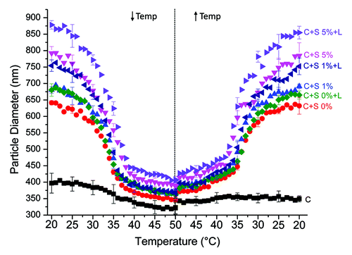
Figure 4. Toxicity of the core + shell nanoparticles in human monocytes. φ, p < 0.05 compared with all other treatments (one-way ANOVA +Tukey post-hoc test). Data are presented as mean ± standard deviation (n = 4).
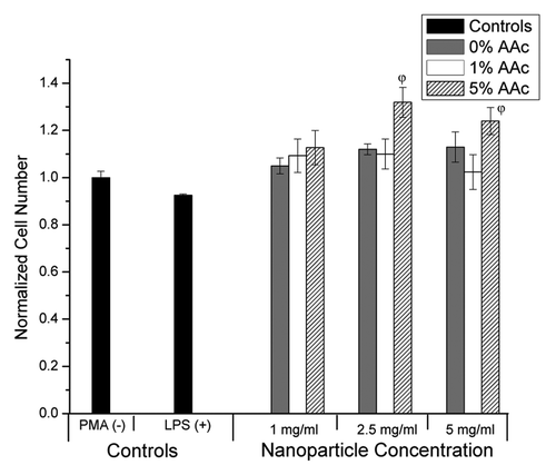
Figure 5. TNF-α production by human monocytes treated with core + shell nanoparticles. φ represents statistical significance from all other data points (p < 0.05, one-way ANOVA + Tukey Post-hoc test). Data presented as mean ± standard deviation (n = 4).
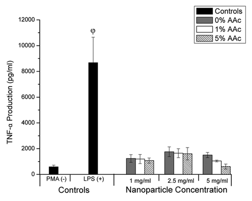
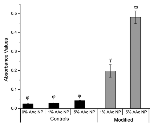
Figure 6. Collagen Type II binding assay for core + shell nanoparticles. 1% and 5% AAc modified NPs are statistically significant from each other and the controls as indicated by the different Greek letters (p < 0.05; one-way ANOVA + Tukey Post-hoc test). Data presented as mean ± standard deviation (n = 4).