Figures & data
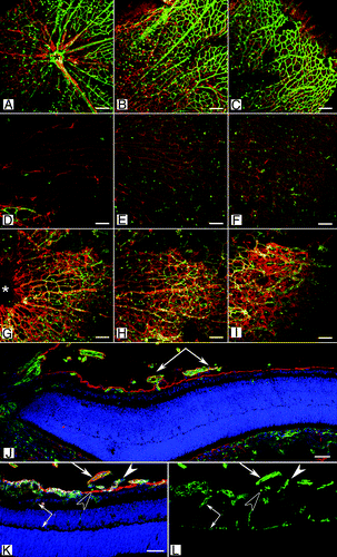
Figure 1. The definition of retinal layers. An adult retinal section stained with hematoxylin and eosin demonstrating the layers of the retina. From the vitreous, labeled on the left side, these are: ILM, inner limiting membrane; GC/NFL, ganglion cell/nerve fiber layer; IPL, inner plexiform layer; INL, inner nuclear layer; OPL, outer plexiform layer; ONL, outer nuclear layer; IPM, interphotoreceptor matrix; BrM Bruch’s membrane (BrM). The laminin chains are shown on the right side where they are expressed in the retina.
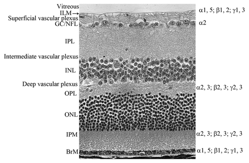
Figure 2. A schematic of retinal vascular development in the normal mouse. Depicted are the key stages in normal vascular development in the mouse. Solid lines indicate blood vessels while broken lines indicate vessels that are regressing. At P1, the hyaloid vasculature in the vitreous (V; white area) consists of the vasa hyloidea proprea (VHP) and the tunica vasculosa lentis (TVL) and the retinal vessels have begun forming. By P7, the superficial vascular plexus in the retina (R) is complete and the TVL and VHP have begun to regress. At P14, the deep retinal plexus is formed and the TVL has regressed. The intermediate plexus forms and the hyaloid vessels completely regressed by P21. The arrow indicates the ILM.
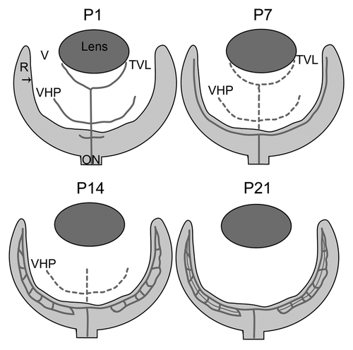
Table1. Laminin chain distribution during retinal development
Figure 3 (See opposite page). Retinal vascular defects in the Lama1 mutant mice. (A–I) Retinal flatmounts from P7 mice were stained with GFAP (red; astrocytes) and GS isolectin (green; blood vessels). In the wild type retina (A–C), vessels and astrocytes spread across the entire retina. Hyaloid vessels (arrows) have begun regressing. By contrast, the retina of Lama1nmf223 mice (D–F) has a vastly reduced number of astrocytes with vessels only in the peripapillary region (D). (A, D, G = peripapillary region; B, E, H = midperiphery; C, F, I = far periphery). When the Lama1 mutants retinas are stained with the hyaloid vessels intact (G–I), astrocytes ensheath these vessels and form a membrane in the vitreous. A cross section from a P7 Lama1 mutant stained with GS isolectin (green) and laminin (red) demonstrates the exit of retinal vessels into the vitreous. Cross sections from P10 Lama1nmf223 eyes labeled with PDGFRα (light blue; astrocytes), GS isolectin (green), anti-pan laminin (red; ILM) and DAPI (blue) show vessels from the vitreous branching into the retina. The inner retinal vessels are forming from these diving vessels (paired arrows). Astrocytes expressing laminin (solid arrowhead) on the vitreal side of the ILM (open arrowhead) near the VHP (arrows). Scale bars indicate (A–I), 100 µm; (J–L), 50 µm. Images in this figure were originally published in BMC Developmental Biology.Citation43